Reflections on impact

In this month’s editorial, we reflect on the journal impact factor and its relationship to impact, especially for publishing methods papers.

Here are a few additional links that readers may find interesting:

The recent HEFCE  (Higher Education Funding Council for England) review of the use of metrics, including the journal impact factor, for research assessment can be found here.

The ASCB’s San Francisco Declaration on Research Assessment (DORA), from a few years ago, can be found here.

An editorial from Nature Materials analyzing the ability of the journal impact factor to predict median citations over 5 years can be found here.

Ten years of Methods

Our tenth anniversary is an occasion to celebrate methods development!

In our Anniversary Issue, we highlight ten areas of methods development, among many candidates, that have had a lot of impact on biological research over the last decade. We also take the opportunity to look back at the papers we have published in some of these areas. We hope to add similar descriptions for all our ‘top-ten methods’ in coming months.

You can look back at the last ten years of Nature Methods in the following areas here:

Microbial sequencing

Super-resolution microscopy

Optogenetics in neuroscience

Light-sheet imaging

Mass-spectrometry based proteomics

High-throughput sequencing data analysis

Anniversary Issue Cover

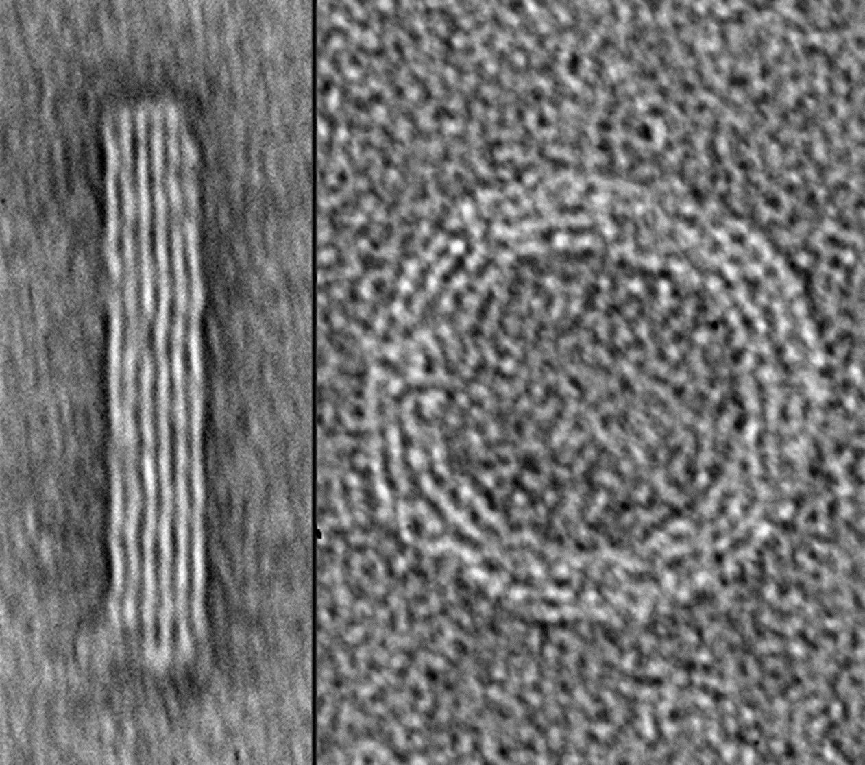
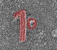
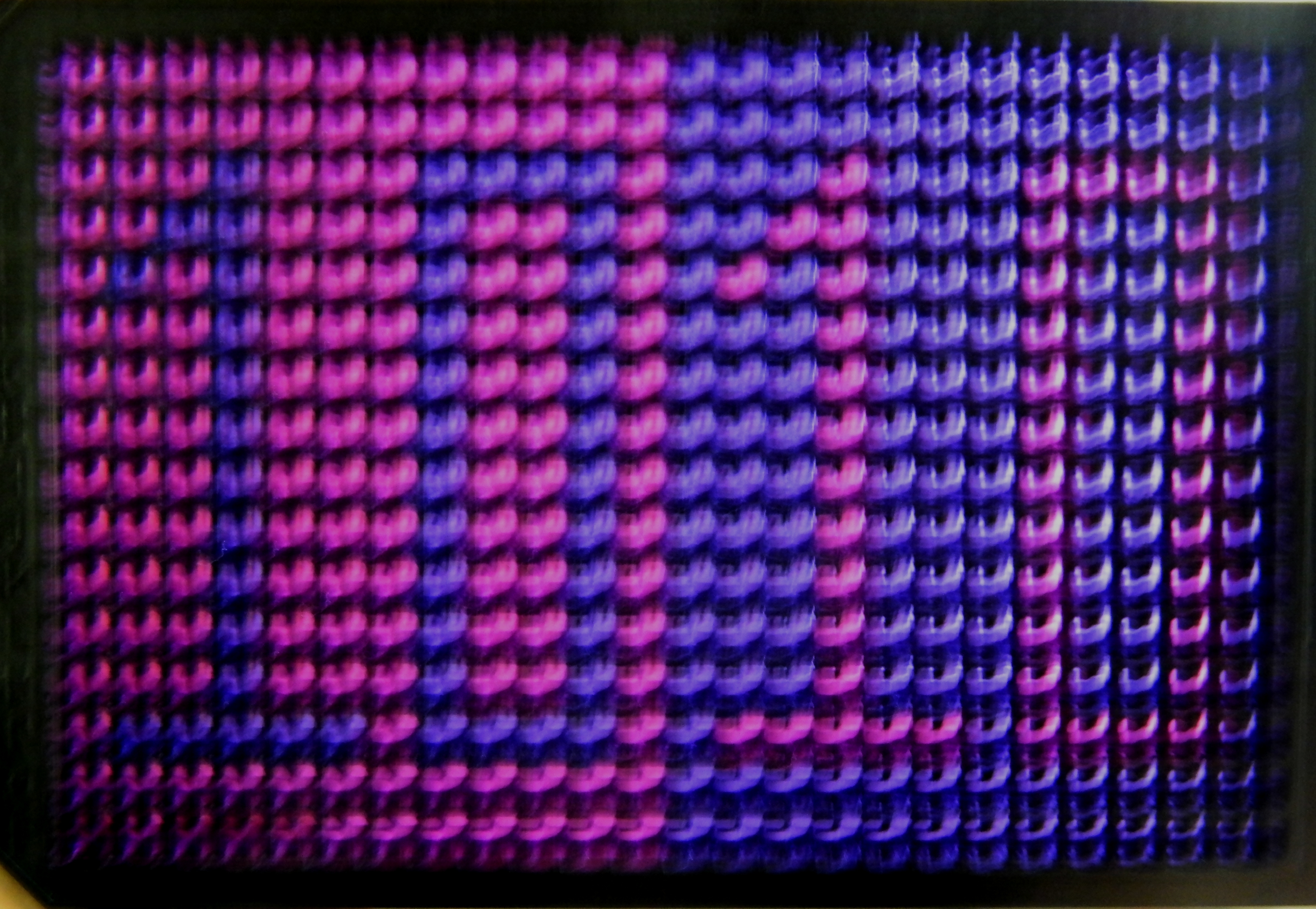
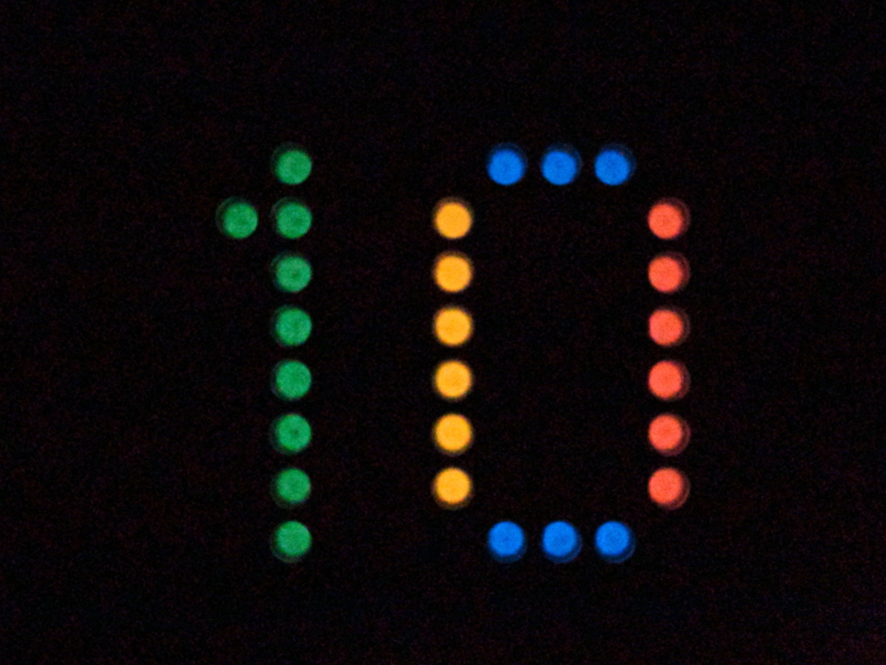
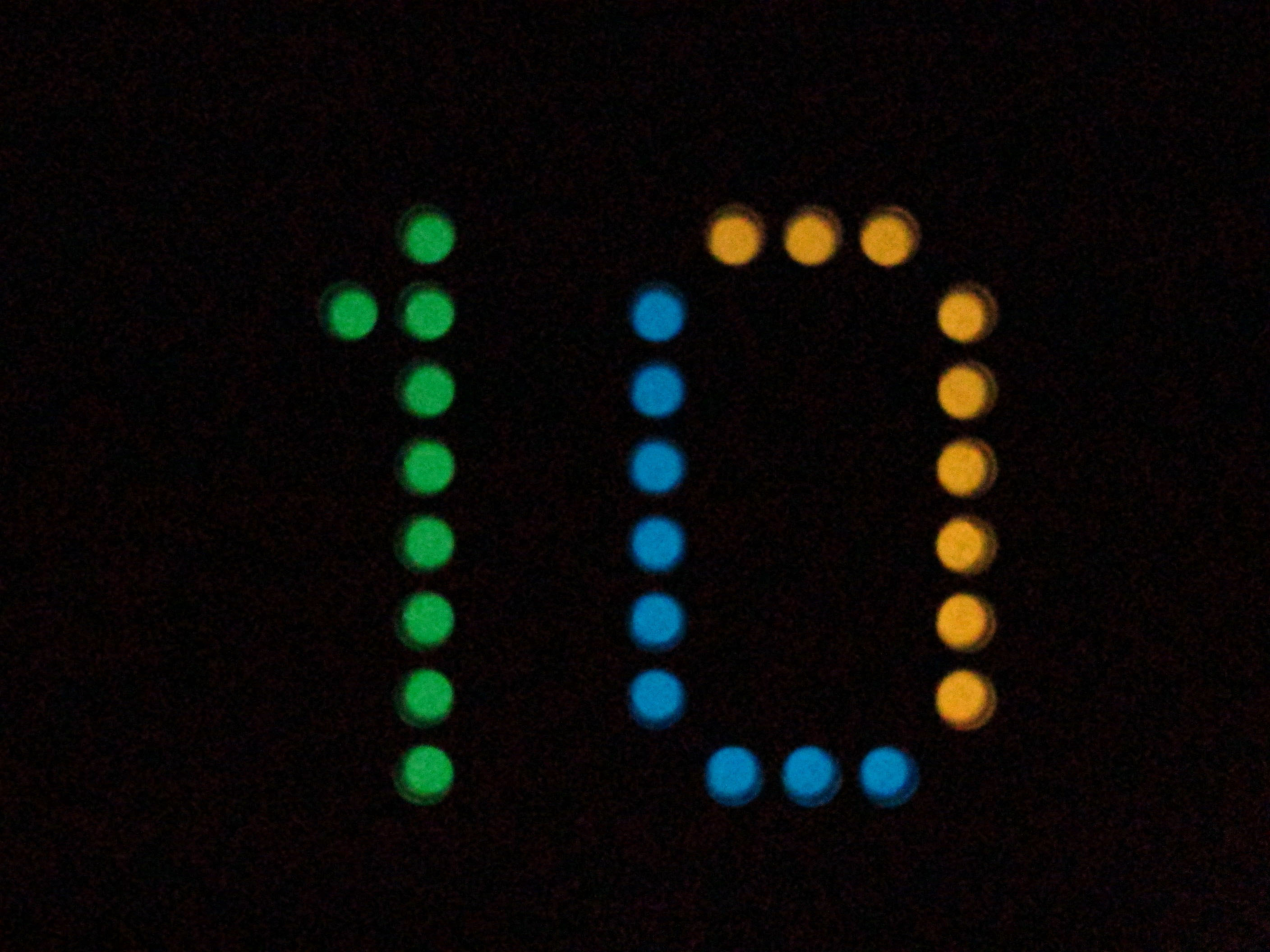
Over the summer we asked for contributions from our readers for the cover of our tenth anniversary issue. We asked for images of the number “10” made using biological research tools and techniques. We were delighted to have many excellent submissions and to be able to use them all on the cover. Here is a bit more detail about these images.

Ke image

{credit}Yonggang Ke{/credit}

Yonggang Ke at Georgia Institute of Technology and Emory University sent us an image of DNA nanostructures. Ke and colleagues used DNA origami to generate two self-assembled 3D nanostructures, imaged them with transmission electron microscopy, and then assembled the images to form the number 10. The height of the final image is 120 nm.


Hogberg cover

{credit}Alan Shaw and Björn Högberg{/credit}

Alan Shaw and Björn Högberg at Karolinska Institutet also applied nanotechnology to the challenge. Building on their recently published Nanocalipers technique (Shaw et al, 2014) they displayed a ferritin protein as the “0” (instead of ephrin as in their published paper) and use DNA origami to generate a nanostructure in the form of a “1”.


DSCN0678-adjusted

{credit}Sandra Duffy{/credit}

Sandra Duffy at Griffith University based her image on indicators of cell viability. Cytotoxic compounds were added to mammalian cells in a 384-well microtiter plate, either in the shape of a 10 in one half of the plate, or to all wells outside the shape of a 10 in the other half of the plate. After incubation, a cell viability marker (resazurin) was added to the wells. Viable cells convert the blue reagent to red, and the image was taken with a simple point-and-shoot camera.


Nano-lantern-2

{credit}Akira Takai, Yasushi Okada, Masahiro Nakano and Takeharu Nagai{/credit}

Nano-lantern-1

{credit}Akira Takai, Yasushi Okada, Masahiro Nakano and Takeharu Nagai{/credit}

Nano-lantern-3

{credit}Akira Takai, Yasushi Okada, Masahiro Nakano and Takeharu Nagai{/credit}

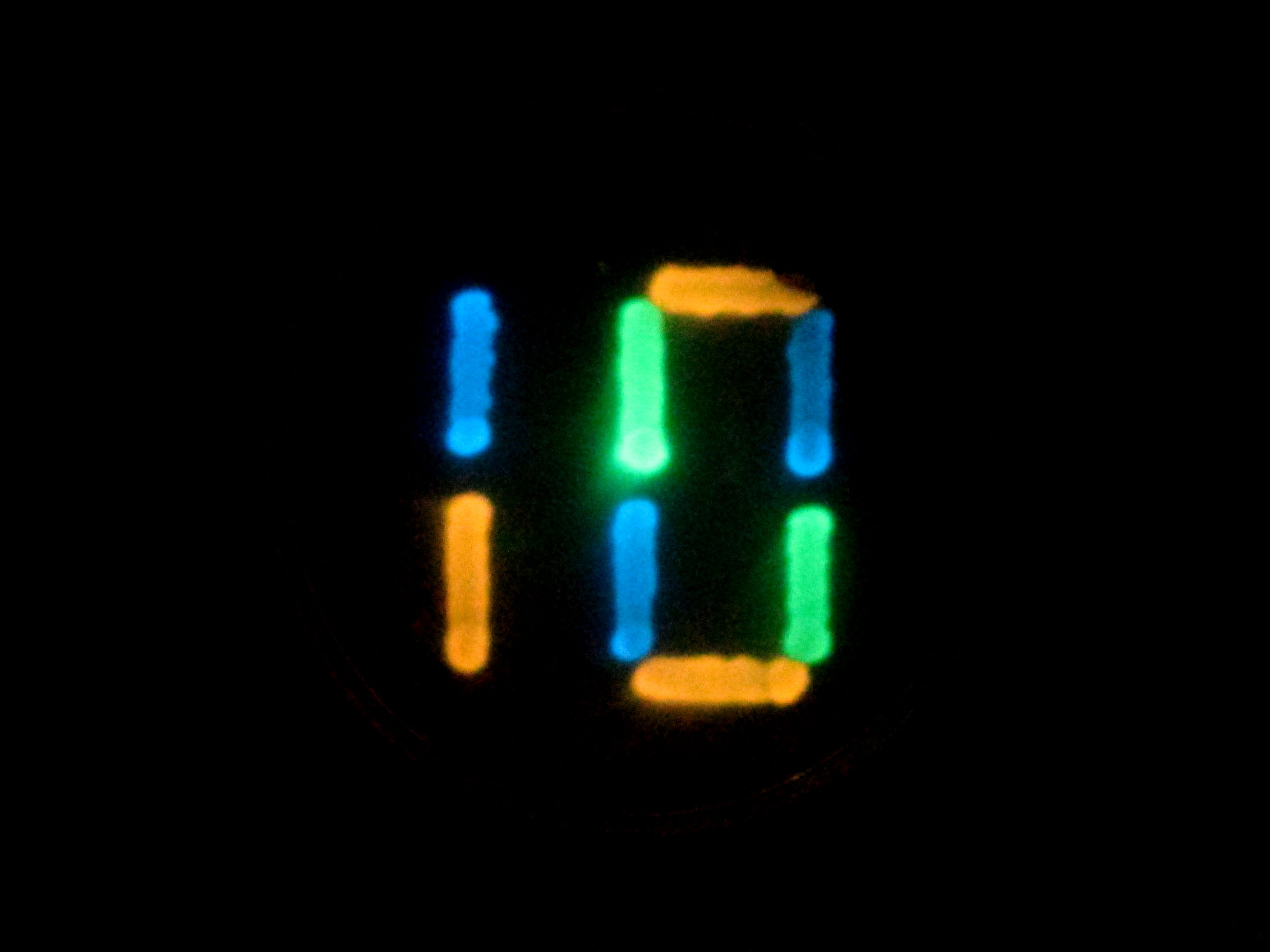
Akira Takai, Yasushi Okada, Masahiro Nakano and Takeharu Nagai, at Osaka University, used multicolour luminescent reporters to write the number 10, either by expressing them in bacterial cells streaked on an agar plate, or by aliquoting them in purified form in a 96-well plate.


10.2-Merge-v2-cropped2

{credit}Lauren Polstein and Charles Gersbach{/credit}

Lauren Polstein and Charles Gersbach at Duke University used light-sensitive transcriptional activators to photoactivate a GFP reporter in mammalian cells in the shape of the number 10.


Navneet Dogra and T. Kyle Vanderlick at Yale University examined bacteria stained with fluorescein (green) interacting with small unilamellar vesicles labeled in red. They used a laser to photobleach all fluorescence except that in the desired shape of the number 10. Image to come.

Finally, Kristina Woodruff and Sebastian Maerkl at EPFL used a standard microarrayer to spot live mammalian cells onto a 675-well array in the shape of a 10 (Woodruff et al, 2013).  Image to come.

We are very grateful to all contributors – thank you for helping us design a cover that salutes the creativity and ingenuity of methods developers!


Optogenetics in neuroscience at Nature Methods

The optogenetic manipulation of cellular properties has not only revolutionized neuroscience, but this technology can also be applied to the manipulation of signaling pathways, transcription or other processes in non-neuronal cells. Here, we highlight some of the papers we have published on the neuroscience side of optogenetics.

Optogenetic tools

2014 has been an exciting year for us with the publication of new optogenetic tools. Klapoetke and Boyden developed Chrimson and Chronos, two channelrhodopsins that they discovered in a screen of algal transcriptomes. Chrimson is more red-shifted than previously known channelrhodopsins while Chronos has faster kinetics. Hochbaum and Cohen described another algal channelrhodopsin called CheRiff, which is highly sensitive to blue light stimulation, making it compatible with red-shifted voltage sensors.

Previously, we published papers describing modifications to optogenetic tools. For example, Prakash and Deisseroth tailored opsin with custom properties. To ensure stoichiometric expression of optogenetic activators and/or inhibitors, Kleinlogel and Bamberg simply and elegantly fused the two proteins into a single chain. Depending on the two partners, this marriage can lead to synergisms or bidirectional effects. Finally, Mattis and Deisseroth undertook a comprehensive characterization of available tools.

Optogenetic applications

Since the initial description of Channelrhodopsin2 (ChR2) as an efficient tool to evoke neural activity in a light-dependent manner, we have seen a flurry of papers applying ChR2 for a variety of questions in neuroscience. For instance, Zhang and Oertner combined this tool with two-photon calcium imaging in rat slices to study synaptic plasticity. Liewald and Gottschalk applied the same methodology to analyze synaptic function in freely moving C. elegans.

ChR2 can also be used to map the function of brains regions as Ayling and Murphy demonstrated by evoking activity in limb muscles via light stimulation in the motor cortex of ChR2 transgenic mice. Similarly, Guo and Ramanathan mapped neural circuitry in C. elegans by combining ChR2-mediated neural activation with imaging of a genetically encoded calcium sensor in downstream neurons. To facilitate circuit mapping in mice, Zhao and Feng generated mouse lines that express ChR2 in GABAergic, cholinergic, serotonergic or parvalbumin-expressing neurons.

While ChR2 is a very popular tool in optogenetics, other family members can do the job as well. C1V1T is a fusion of two different opsins and is particularly useful when applying two-photon excitation, as shown by Packer and Yuste. ReaChR is activated by red light and thus especially useful in vivo. Inagaki and Anderson studied courtship behavior in Drosophila with this tool.

Method of the Year

We celebrated the impact of optogenetics by recognizing the technology as our Method of the Year 2010. We marked the occasion with the publication of special Commentaries on the subjects. Deisseroth discussed the past, present and future of optogenetics. Hegemann and Möglich deliberate on the exploration of new optogenetic tools. And Peron and Svoboda illuminated us on the precise delivery of optogenetic stimulation. In addition, our News Feature recounted the stories behind the “Light tools”.

If we have sparked your interest, the mentioned papers are listed below.

We are excited to hear about the upcoming developments in optogenetics from you.


Nathan C Klapoetke, Yasunobu Murata, Sung Soo Kim, Stefan R Pulver, Amanda Birdsey-Benson, Yong Ku Cho, Tania K Morimoto, Amy S Chuong, Eric J Carpenter, Zhijian Tian, Jun Wang, Yinlong Xie, Zhixiang Yan, Yong Zhang, Brian Y Chow, Barbara Surek, Michael Melkonian, Vivek Jayaraman, Martha Constantine-Paton, Gane Ka-Shu Wong & Edward S Boyden
Independent optical excitation of distinct neural populations
Nature Methods 11, 338–346 (2014) doi:10.1038/nmeth.2836

Daniel R Hochbaum, Yongxin Zhao, Samouil L Farhi, Nathan Klapoetke, Christopher A Werley, Vikrant Kapoor, Peng Zou, Joel M Kralj, Dougal Maclaurin, Niklas Smedemark-Margulies, Jessica L Saulnier, Gabriella L Boulting, Christoph Straub, Yong Ku Cho, Michael Melkonian, Gane Ka-Shu Wong, D Jed Harrison, Venkatesh N Murthy, Bernardo L Sabatini, Edward S Boyden, Robert E Campbell & Adam E Cohen
All-optical electrophysiology in mammalian neurons using engineered microbial rhodopsins
Nature Methods 11, 825–833 (2014) doi:10.1038/nmeth.3000

Rohit Prakash, Ofer Yizhar, Benjamin Grewe, Charu Ramakrishnan, Nancy Wang, Inbal Goshen, Adam M Packer, Darcy S Peterka, Rafael Yuste, Mark J Schnitzer & Karl Deisseroth
Two-photon optogenetic toolbox for fast inhibition, excitation and bistable modulation
Nature Methods 9, 1171–1179 (2012) doi:10.1038/nmeth.2215

Sonja Kleinlogel, Ulrich Terpitz, Barbara Legrum, Deniz Gökbuget, Edward S Boyden, Christian Bamann, Phillip G Wood & Ernst Bamberg
A gene-fusion strategy for stoichiometric and co-localized expression of light-gated membrane proteins
Nature Methods 8, 1083–1088 (2011) doi:10.1038/nmeth.1766

Joanna Mattis, Kay M Tye, Emily A Ferenczi, Charu Ramakrishnan, Daniel J O’Shea, Rohit Prakash, Lisa A Gunaydin, Minsuk Hyun, Lief E Fenno, Viviana Gradinaru, Ofer Yizhar & Karl Deisseroth
Principles for applying optogenetic tools derived from direct comparative analysis of microbial opsins
Nature Methods 9, 159–172 (2012) doi:10.1038/nmeth.1808

Yan-Ping Zhang & Thomas G Oertner
Optical induction of synaptic plasticity using a light-sensitive channel
Nature Methods 4, 139 – 141 (2006) doi:10.1038/nmeth988

Jana F Liewald, Martin Brauner, Greg J Stephens, Magali Bouhours, Christian Schultheis, Mei Zhen & Alexander Gottschalk
Optogenetic analysis of synaptic function
Nature Methods 5, 895 – 902 (2008) doi:10.1038/nmeth.1252

Oliver G S Ayling, Thomas C Harrison, Jamie D Boyd, Alexander Goroshkov & Timothy H Murphy
Automated light-based mapping of motor cortex by photoactivation of channelrhodopsin-2 transgenic mice
Nature Methods 6, 219 – 224 (2009) doi:10.1038/nmeth.1303

Zengcai V Guo, Anne C Hart & Sharad Ramanathan
Optical interrogation of neural circuits in Caenorhabditis elegans
Nature Methods 6, 891 – 896 (2009) doi:10.1038/nmeth.1397

Shengli Zhao, Jonathan T Ting, Hisham E Atallah, Li Qiu, Jie Tan, Bernd Gloss, George J Augustine, Karl Deisseroth, Minmin Luo, Ann M Graybiel & Guoping Feng
Cell type–specific channelrhodopsin-2 transgenic mice for optogenetic dissection of neural circuitry function
Nature Methods 8, 745-752 (2011) doi:10.1038/nmeth.1668

Adam M Packer, Darcy S Peterka, Jan J Hirtz, Rohit Prakash, Karl Deisseroth & Rafael Yuste
Two-photon optogenetics of dendritic spines and neural circuits
Nature Methods 9, 1202–1205 (2012) doi:10.1038/nmeth.2249

Hidehiko K Inagaki, Yonil Jung, Eric D Hoopfer, Allan M Wong, Neeli Mishra, John Y Lin, Roger Y Tsien & David J Anderson
Optogenetic control of Drosophila using a red-shifted channelrhodopsin reveals experience-dependent influences on courtship
Nature Methods 11, 325–332 (2014) doi:10.1038/nmeth.2765

Karl Deisseroth
Optogenetics
Nature Methods 8, 26–29 (2011) doi:10.1038/nmeth.f.324

Peter Hegemann & Andreas Möglich
Channelrhodopsin engineering and exploration of new optogenetic tools
Nature Methods 8, 39–42 (2011) doi:10.1038/nmeth.f.327

Simon Peron & Karel Svoboda
From cudgel to scalpel: toward precise neural control with optogenetics
Nature Methods 8, 30–34 (2011) doi:10.1038/nmeth.f.325

Monya Baker
Light tools
Nature Methods 8, 19–22 (2011) doi:10.1038/nmeth.f.322

Light sheet imaging in Nature Methods

It was only a few months before Nature Methods was launched in October 2004 that Jan Huisken and Ernst Stelzer had published a paper in Science in which they used light sheet microscopy – what they called selective plane illumination microscopy or SPIM – to image fluorescence within transgenic embryos. Simplistically put, this century-old technique achieves optical sectioning by illuminating a sample through its width with a thin sheet of light. In the last decade, Nature Methods has published a steady stream of papers reporting developments in light-sheet imaging. Here are the highlights.

Our very first light-sheet paper was also from the Stelzer group, reporting the use of deconvolution to improve resolution of the technique (Verveer et al, 2007). This was rapidly followed by a paper from Hans-Ulrich Dodt, in which samples such as entire insects or brain tissue were rendered transparent with clearing agents to produce spectacular light-sheet ‘ultramicrographs’ (Dodt et al, 2007). The push to higher resolution continued, with a paper from Albert Diaspro reporting 3D super-resolution imaging within thick samples using light-sheets (Cella Zanacchi et al, 2011). The Stelzer group, meanwhile, improved performance of the technique in larger samples that scatter more light, by combining it with structured illumination (Keller et al, 2010).

Thai Truong, Willy Supatto and Scott Fraser added two photon excitation to light-sheet imaging, thereby doubling the depth and increasing by an order of magnitude the speed at which they could image samples such as developing embryos with each approach alone (Truong et al, 2011); Supatto recently extended this to imaging in multiple colours  (Mahou et al, 2014). And then in 2012, the groups of Phillip Keller and Lars Hufnagel independently reported microscopes that could take take multiple views of a biological sample simultaneously, allowing rapid imaging of entire developing fly embryos at sub-cellular resolution (Tomer et al, 2012; Krzic et al, 2012).

Though light-sheet imaging is perhaps at its most powerful in the imaging of thick samples like embryos or tissue sections, it has been used for substantial performance improvements in cellular imaging as well. In 2011, Eric Betzig’s group used scanned Bessel beams to create thinner light sheets and thus much improved axial resolution, achieving isotropic 3D resolution and rapid imaging within living cells (Planchon et al, 2011). Note also that, as Tom Vettenburg, Kishan Dholakia and colleagues showed,  generating the light sheet using an Airy beam, rather than Gaussian or Bessel beam, yields an even larger field of view without sacrificing contrast and resolution (Vettenburg et al, 2014). Variations on the light-sheet theme have also been developed by the labs of Makio Tokunaga and Sunney Xie for single-molecule imaging within cells (Tokunaga et al, 2008; Gebhardt et al, 2013).

In recent years, the excitement around this technology has been palpable, with several papers reporting impressive applications of light-sheet microscopy: it has been used to functionally image the entire fish brain (Ahrens et al, 2013) and the brain of ‘fictively behaving’ fish (Vladimirov et al, 2014), as well as to image the beating fish heart (Mickoleit et al, 2014).

Perhaps not surprisingly, the emphasis in methods development has also been shifting a little. On the one hand, platforms are being developed to make this valuable technique available more widely, for instance via the OpenSPIM or OpenSpinMicroscopy platforms (Pitrone et al, 2013; Gualda et al, 2013). At the same time, analytical tools are necessarily being developed to handle the vast reams of data that a light-sheet experiment generates. The group of Pavel Tomancak reported Bayesian-based deconvolution methods to analyse the large data sets that result from multiview imaging (Preibisch et al, 2014). Phillip Keller and colleagues described computational methods to segment and track nuclei in data sets from light sheet or other imaging, for fast lineaging of developing embryos (Amat et al, 2014). Misha Ahrens and colleagues reported Thunder, a suite of analytical tools built on a platform for distributed computing, enabling the mapping of brain activity in ‘fictively behaving’ zebrafish (Freeman et al, 2014).

It’s fair to say that this venerable method has been thoroughly revived over the past decade. Light-sheet imaging is poised to yield tremendous biological insight. We hope to keep you updated on future developments in Methagora.