
Not CAT scans; actual cats.
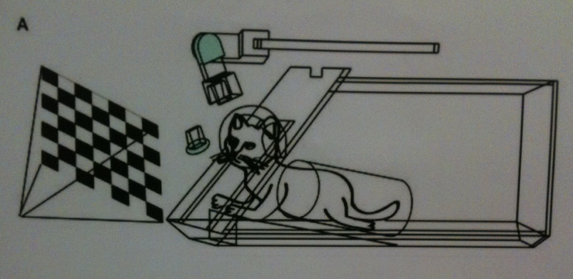
{credit}Manxui Ma{/credit}
Faced with a difficult task, people will often compare their situation to ‘herding cats’. I can only imagine the degree of herding that had to go on in a study reported in a poster yesterday here at the meeting of the OHBM (Organization for Human Brain Mapping).
Most people using fMRI (functional magnetic resonance imaging), a non-invasive way of looking at brain activity, like to study humans. But scanning animals is also helpful because you can use stronger magnets to get really detailed pictures of brain networks; it’s also helpful for comparing findings across species, or matching up the decades of work on animals to the results being churned out of fMRI scanners today.
One problem is that any movement of the head once your subject is in the scanner can really mess up your signal. So scientists usually anaesthetize animals before they are scanned. The only animals that have been scanned while awake are monkeys and dogs, mainly because you can train them fairly effectively to keep still. Now, Manxiu Ma and colleagues from the Human Brain Mapping Center at the Chinese Academy of Sciences in Beijing have tried it with two cats.
Why cats? Well, this is really a proof-of-concept — but plenty of anatomical work has been done in cats to looking at the effects on the brain of changing input — covering one eye from birth, for example, and watching to see how the visual cortex changes. So they’re already a model in neuroscience. In addition, they’re small, which means that you can use comparatively stronger magnets and see the brain in more detail. With human scanners there’s a trade-off between size and power. Continue reading
