EMBO Young Investigators

This month saw the announcement of the EMBO Young Investigators for 2014, and we’re very pleased to count three of them amongst our Nature Protocols Authors! All in all, 27 researchers from 11 different countries were selected to receive the status of EMBO Young Investigator this year; you can see the full list of researchers along with their research interests here.

So, we’d like to congratulate all the new EMBO Young Investigators, and especially our Authors Filippo Del Bene, Ines Thiele and Johannes Zuber. Ines has written two Nature Protocols on the topic of metabolism, one for generating genome-scale metabolic reconstructions and a second for predicting cellular metabolism using the COBRA toolbox, both of which have been very popular additions to our content. With a focus on RNAi, Johannes’ protocol provides a pipeline for the generation of shRNA transgenic mice, and Filippo’s protocol for converting zebrafish transgenic lines from eGFP reporters to GAL4 drivers with CRISPR/Cas9 is one of our most recent articles, published in Nature Protocols earlier this month. It’s great that we’ve been able to help disseminate the work of such up-and-coming researchers, and we’re equally pleased that they have found us the place to publish their protocols!

Notes from the Single Cell Analysis Europe conference

At the end of March, Dot and I spent two days in Edinburgh at the Select Biosciences meetings held at the Edinburgh Conference Centre on the Herriot-Watt campus, about 6 miles outside the city centre. The weather was record-breakingly hot for March (and Scotland!) – here is some evidence of the spring sunshine:


We didn’t have too much time to enjoy the weather though as there were four concurrent meetings to attend; I spent almost all of my time at the Single Cell Analysis Europe conference, while you will receive further reports from Dot on talks from the Lab-on-a-chip European Congress, Advances in Microarray Technology, and Advances in Biodetection & Biosensors sessions.

As an editor, I found the meeting really interesting, with some of the top names in the field speaking and methodological advances a-plenty for us to learn about!  The exhibition hall was also a really interesting addition to the main sessions as we got to see a lot of the lab-on-a-chip and microfluidic equipment that people use in their protocols in real life for the first time! And they really are quite amazing!

Day one’s session on using microarrays and chips in single cell analysis started with a keynote presentation from Professor Marcus Textor from ETH Zurich, on the use of microfabricated cell culture platforms to study cell function and drug response in engineered 2D and 3D microenvironments. Describing how cell culture has moved on from the invention of the Petri dish in 1877, Professor Textor firstly showed how PDMS microwells can be engineered to provide 3D culture environments with varying substrate stiffness and shape, which can regulate the assembly of the actin cytoskeleton, impacting cell survival, phenotype and function. He also described the use of a PEG-based microwell platform to create 3D breast cancer models to allow responses to taxol and other anticancer drugs to be tested in vitro.

Dr. John Collins (NanoInk Inc.) then presented a talk on dip pen nanolithography as a technique for fabricating cellular microenvironments that allows single cell co-culture, and targeted delivery of agents such as drugs. As a work in progress, Dr. Collins also explained how they are using this technology to print substrates in lines rather than dots to enable the study of cell motility in this 3D microenvironment.

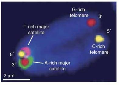
The next session, on label-free routes to single cell analysis started with an interesting talk from Professor Susann Müller (Helmholtz Centre of Environmental Research), which focused on assessing microbial community dynamics using flow cytometry and phylogenetics, encompassing analysis of even unculturable bacterial strains. Changes in the community cytometric fingerprint or structure over time can be measured and used as a biosensor for the state of natural environments, such as monitoring the stability of the enhanced biological phosphorous removal (EBPR) process in wastewater treatment plants as illustrated in this talk.

The afternoon began with the second keynote presentation, from Professor Nicholas Navin of the MD Anderson Cancer Center, which detailed the investigation of genome evolution in breast cancer by single cell sequencing. Combining flow-cytometric sorting, whole genome amplification and next-generation sequencing, this single nucleus sequencing (SNS) method can be used to accurately quantify copy number in single nuclei. Professor Navin’s group used SNS to analyse 100 single cells from a polygenomic tumour and 100 single cells from a monogenomic primary tumor and its liver metastasis, data from which indicates that tumors grow and evolve by punctuated clonal expansions, rather than gradual tumor progression. Look out for the upcoming Nature protocol on this method as we have this in production as we speak!

New and improved methods for DNA sequencing are clearly an up-and-coming area of methodological research and before moving on to the use of microfluidics in single cell analysis, the afternoon’s session continued with a talk from Professor David Bensimon (Ecole Normale Supérieure) describing a novel, single molecule method for the mechanical sequencing of DNA. This method uses a magnetic trap to mechanically open out and close DNA hairpins. In the presence of hybridizing complementary oligonucleotides, rehybridization of DNA hairpins is blocked, and the position of these roadblocks on single molecules can be measured with nanometer precision, allowing sequencing of the DNA molecule by hybridization or ligation: 

For further details on this new method, see the paper just published in this issue of Nature Methods (9(4), p367-372) [Update: this paper is now free to access for a week] and the News & Views piece by Sten Linnarsson in the same issue, from which the above illustration is taken. Another method for sequencing was also presented at the same time in the lab-on-a-chip meeting, using nanopore sensors for next generation DNA sequencing. Sadly my conference-going skills do not extend to splitting myself in half to be at two talks at once, so I was unable to hear about this new method in person as well!

Day two started with two talks on the use of RT-qPCR in single cell analysis, from Professor Mikael Kubista (TATAA Biocenter) and Dr. Ken Livak (Fluidigm). These highlighted the variation in gene expression levels found from cell-to-cell, even in seemingly homogeneous populations, caused by the stochastic nature of transcription that occurs in the cell in bursts. Both speakers presented methods that can be used to deal with this heterogeneity, the latter explaining why single cell data needs to be treated differently from conventional qPCR data, and how this can be achieved using multivariate analysis and considering factors such as replicates, limits of detection, normalization and data display.

A couple more very interesting methodological talks followed in the afternoon session on single cell analysis in signalling, from Professor Ola Soderberg (Uppsala University) and Frederik Fritzsch (Dortmund University). Professsor Soderberg described the application of the in situ proximity ligation assay (PLA) and padlock probes to visualize signal pathway activity in single cells. In situ PLA utilizes pairs of antibodies, with bound DNA sequences, to target interacting proteins. Proximal binding of the antibodies and the conjugated oligonucleotide sequences creates a circular DNA molecule that can be amplified by rolling circle amplification (RCA) and subsequently detected by hybridization of fluorophore-labelled probes. Padlock probes also use RCA to generate signals for detection and the two can be combined to allow multiplexed analyses. The Envirostat 2.0 was then presented by Frederik Fritzsch with plenty of impressive videos to demonstrate this new, negative dielectrophoresis-based system for contactless single cell isolation, cultivation and analysis.

All in all, the full program provided many stimulating talks in areas undergoing, and with great potential for, methods development. Although we cover protocols in these areas already, we are now full of new ideas for enhancing our content on these topics going forward, so watch this space!

Single Cell Analysis Europe

Next week, Dot and I will be heading to Edinburgh to attend the Select Biosciences Single Cell Analysis Europe conference.  These days, the analysis of single cells is being increasingly applied to get to the bottom of experimental questions in biology, and is a rapidly expanding discipline, rich in methodological advances.  We are really looking forward to hearing about the techniques currently being used, as well as discovering new methods that are now being developed.

The conference will run over two days, 28th-29th March, and covers the use of microarrays, microfluidics and label-free approaches to study single cells, as well as the use of RT-qPCR in single cell analysis, among other topics.  Keynote lectures will be given by Norman Dovichi and Marcus Textor, and two of our very own Nature Protocols authors, Jay Groves and Nicholas Navin.  Although our single nucleus sequencing protocol from the latter is only just in production at the moment, we have a Nature protocol from the Groves lab. that describes the use of supported membranes and single-cell microscopy to investigate cell signalling. We also have a Nature protocol on RT-PCR data analysis from Kenneth Livak, which is relevant to his upcoming talk at this conference, and a Nature protocol from Susann Müller who will be speaking there too, describing how to use microbial community dynamics as a biosensor.

In advance of our attendance, we have put together a collection of our single-cell protocols for your interest, which will be free to access for the rest of this month, alongside those from our authors who will be speaking.  While this is only an example of our content on this topic, you will see that the protocols cover a range of areas from gene expression and copy number profiling to isolation and imaging of single cells and we hope that you will find them useful!

In addition, conference registration also allows entrance to three other concurrent meetings; the Lab-on-a-chip European Congress, Advances in Microarray Technology, and Advances in Biodetection & BiosensorsNature Protocols authors are again represented amongst the speakers at these meetings, with Mark Bradley (protocol in production), Andreu Llobera and Bo Mattiasson all giving talks.  As you can see, these meetings are also highly relevant to our scope and content at Nature Protocols so we are keen to make it to some of these sesions too.  There are only two of us, but we will aim to get to as many of the talks as we can and will bring you some reports shortly after our return!


Notes from the 3rd EMBO meeting, Vienna.

So I have finally finished collecting my thoughts from the 3rd EMBO meeting in Vienna, which I was fortunate enough to attend from the 10th-13th Sept.

Hosted in the Austria Centre Vienna

Austria Centre Vienna

the meeting was attended by around 1300 people, and with over 600 posters and 21 concurrent sessions there was lots to keep participants busy! As my first conference experience for a good few years, it was a little overwhelming, but in a good way!

Vienna

I found Vienna to be a lovely city, as was the weather for the weekend, which helps with the laid back atmosphere…

MuseumsQuartier

The architecture is amazing, with beautiful buildings on every corner

Karlskirche

and although it was a little too cultural for my time-frame with many art galleries and museums that I didn’t manage to see, I did appreciate my visit to see the living art of the Spanish Riding School! Sadly we weren’t allowed to take pictures of the action, but here is one of the stars dozing in the sun

Lipizzaner

Anyway, on to the conference, which began with a series of plenary lectures on hosts & microbes, including a really interesting talk by Bonnie Bassler who explained their work investigating quorum sensing inhibitors as potential therapeutics to prevent pathogenesis. Although there is still some way to go before inhibitor compounds are made into drugs, this sounds like an exciting avenue of future research, especially given the multidrug resistance problems nowadays. I also really enjoyed the final talk in this session from Brett Finlay (despite the high content of toilet humour!) on the importance of our gut microbiota in our health and their role in resistance to allergic diseases such as asthma.

For the next 3 days, there was a great diversity of topics presented so I was too often spoilt for choice about which session to attend! Day two started with an excellent keynote lecture from Susan Lindquist discussing the role of protein folding in the inheritance of environmentally acquired traits via both Hsp90- and prion-mediated mechanisms. She explained how Hsp90 buffers the effects of genetic variation and acts as a potentiator of evolution, enabling previously hidden genetic variation to be revealed in times of stress. In the plenary session on genome evolution that followed, Paul Rainey presented evidence of the involvement of an epigenetic switch in bacterial genome evolution, reinforcing the theme that genome evolution is about so much more than just the underlying genetic variation.

Next-generation sequencing

Another theme that spanned many sessions was the use of next-generation sequencing and the advances made possible in a variety of fields as a result of the development of these techniques. This was illustrated by Mike Stratton’s plenary lecture on the exome sequencing and analysis of cancer genomes. Hand-in-hand with such technological advances of course comes the need for methods to analyse the vast quantities of data produced. These ideas were further explored in the session on human genome variation & disease with a talk from Elaine Mardis covering whole genome sequencing approaches used to analyse cancer genomes, including their application to personal diagnosis and treatment.

Personal medicine

Continuing on the topic of personal medicine, Russ Altman from Stanford next introduced a pharmacogenomics database designed to allow the effect of human genetic variation on drug responses to be assessed. This became quite entertaining when he reported results from the analysis of the personal genome of his colleague, Steve Quake; he had obviously agreed to everyone at EMBO and beyond knowing how he is likely to respond to a variety of drugs as well as his genetic predisposition to various diseases, but it is quite wierd to feel like I know far more about his genome than about the man himself?!

Computer vision

As well as the increase in the throughput of sequencing methods, there is also now a need for computational methods to keep up with advances in imaging methods. This was explored in a stimulating session on computer vision of cellular function in which Andre Brown and Sean Megason (amongst others) described systems to image C. elegans behaviour and zebrafish embryo development, respectively, as well as outlining the software they designed to analyse the image data obtained. The real take home message from this session was that it’s no use having the means to obtain data on this scale if you don’t also have algorithms and software to enable efficient and accurate analysis of this data to extract the biological meaning. So I think this will be a big area for methods development going forwards.

TNTs

I also found a new area of molecular biology to me and was really interested to learn about the recent discovery of tunneling nanotubes (or TNTs). In a talk by Hans-Hermann Gerdes we heard how these nanotubes form and that they are used for long distance electrical coupling between cells, and as a method of cell-cell communication for the transfer of cytoplasmic molecules and organelles. Fascinating stuff!

And finally, the prize for the best talk goes to…

Florian Engert, for his virtual reality world for zebrafish larvae. Or as he put it, ‘The Matrix’ for fish! He presented work on adaptive motorlearning in zebrafish larvae and detailed the system he developed in which larvae are paralyzed and surrounded by a virtual reality environment, which allows the whole brain to be imaged at a single cell level in an awake animal as they respond to experimentally induced changes in the environment. This clearly provides an exciting system to study the neural circuitry underlying a range of visually-induced behaviours in zebrafish.

Featured protocols

I will finish with a reminder of a couple of Nature Protocols that I was really pleased to spot being put to good use in the work presented at EMBO.

Firstly, “CO-FISH “:https://www.nature.com/nprot/journal/v5/n7/full/nprot.2010.102.html (chromosome orientation fluorescent in situ hybridization) was being utilised by both Shahragim Tajbakhsh and Peter Lansdorp in their talks on the non-random segregation of chromosomes in mitosis.

nprot_2010_102-F1.jpg

And genomic SELEX formed the basis of the work presented in Renee Schroeder’s talk in the RNA & RNA mediated regulation session.

nprot_2006_372-F1.jpg

The arrival of guidelines for reporting experiments

Are you conducting experiments using animals or developing protocols in this area of research? If so, you may be interested to take a look at the ARRIVE (Animal Research: Reporting In Vivo Experiments) guidelines when reporting your results and writing up those experimental procedures. These guidelines were published in June 2010 to improve the reporting of research using animals and can be found here. The guidelines themselves are presented in Table 2 of this paper, and consist of twenty items that can be used as a checklist for authors preparing a manuscript on this topic. Of course they are useful for peer-reviewers and journal editors too!

The ARRIVE guidelines provide recommendations about the content of all the standard sections of a manuscript, covering many points you would expect such as ethics statements and description of the experimental procedures and animals used. In addition, they suggest how to report the statistical analysis of results, as well as providing information about the study design, which is particularly relevant when writing up a protocol. To apply these guidelines to the preparation of a protocol for Nature Protocols, an Experimental Design section in the Introduction can be a good place to include further information on study design and the housing and husbandry of experimental animals, while the Procedure and Anticipated Results sections obviously allow detailed description of the methods used to obtain and analyse the results.

Although we do not publish primary research at Nature Protocols, but instead focus on tried-and-tested methods that have been previously published, we do find that these guidelines provide a useful resource and we encourage our authors to comply with the ARRIVE reporting guidelines when documenting protocols that involve animal studies.