Anniversary Issue Cover

Over the summer we asked for contributions from our readers for the cover of our tenth anniversary issue. We asked for images of the number “10” made using biological research tools and techniques. We were delighted to have many excellent submissions and to be able to use them all on the cover. Here is a bit more detail about these images.

Ke image

{credit}Yonggang Ke{/credit}

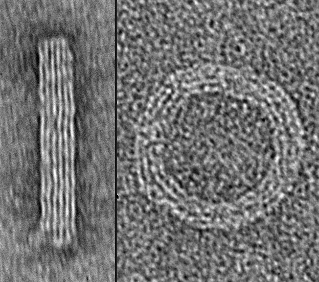
Yonggang Ke at Georgia Institute of Technology and Emory University sent us an image of DNA nanostructures. Ke and colleagues used DNA origami to generate two self-assembled 3D nanostructures, imaged them with transmission electron microscopy, and then assembled the images to form the number 10. The height of the final image is 120 nm.


Hogberg cover

{credit}Alan Shaw and Björn Högberg{/credit}

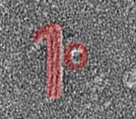
Alan Shaw and Björn Högberg at Karolinska Institutet also applied nanotechnology to the challenge. Building on their recently published Nanocalipers technique (Shaw et al, 2014) they displayed a ferritin protein as the “0” (instead of ephrin as in their published paper) and use DNA origami to generate a nanostructure in the form of a “1”.


DSCN0678-adjusted

{credit}Sandra Duffy{/credit}

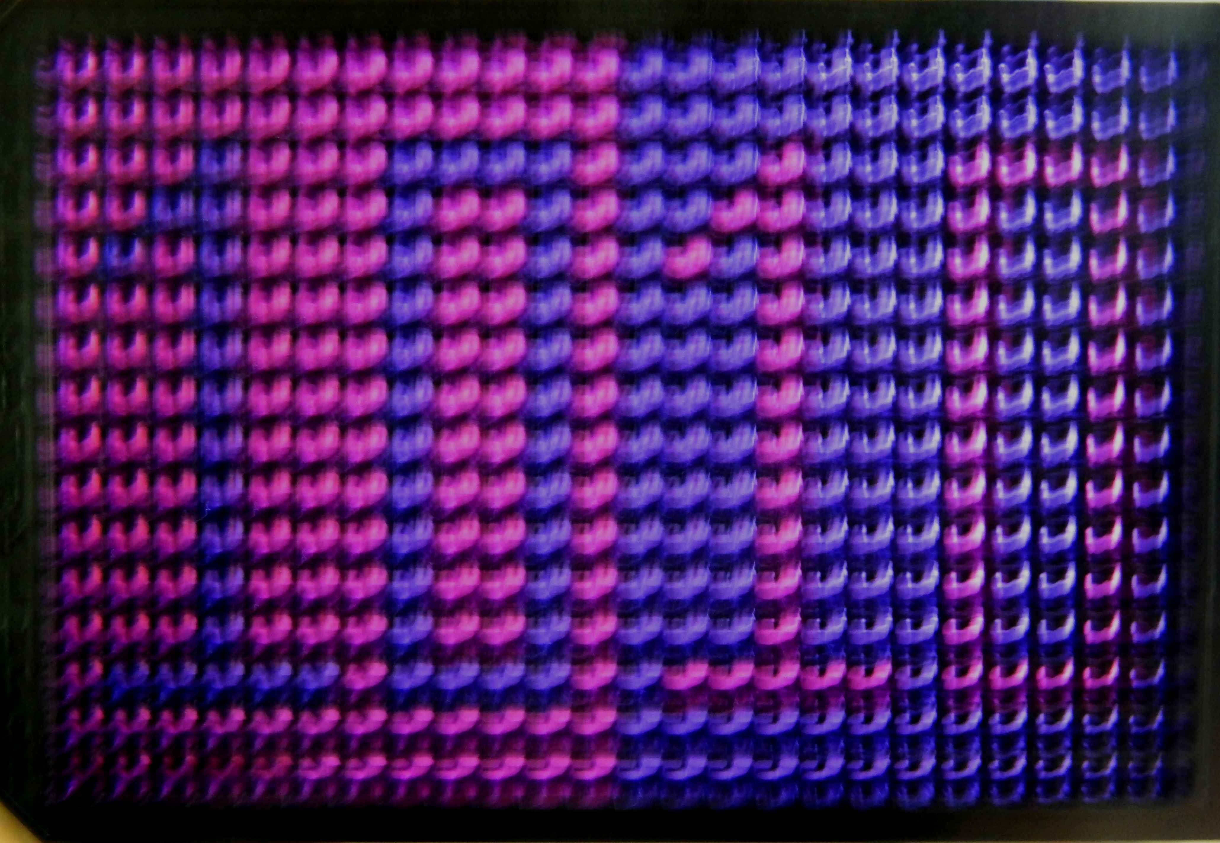
Sandra Duffy at Griffith University based her image on indicators of cell viability. Cytotoxic compounds were added to mammalian cells in a 384-well microtiter plate, either in the shape of a 10 in one half of the plate, or to all wells outside the shape of a 10 in the other half of the plate. After incubation, a cell viability marker (resazurin) was added to the wells. Viable cells convert the blue reagent to red, and the image was taken with a simple point-and-shoot camera.


Nano-lantern-2

{credit}Akira Takai, Yasushi Okada, Masahiro Nakano and Takeharu Nagai{/credit}

Nano-lantern-1

{credit}Akira Takai, Yasushi Okada, Masahiro Nakano and Takeharu Nagai{/credit}

Nano-lantern-3

{credit}Akira Takai, Yasushi Okada, Masahiro Nakano and Takeharu Nagai{/credit}

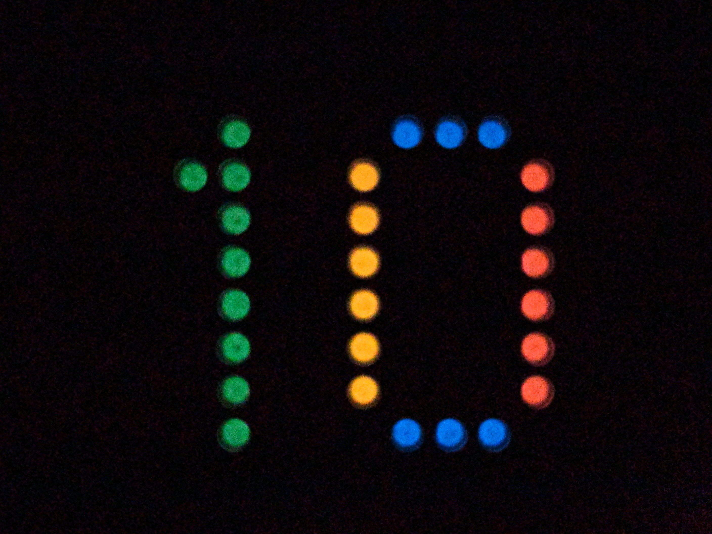
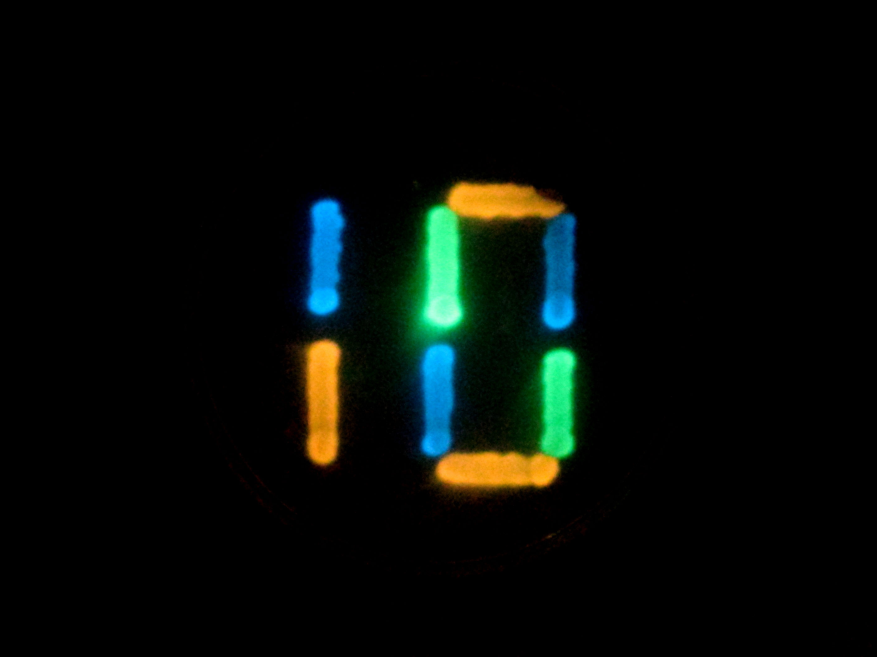
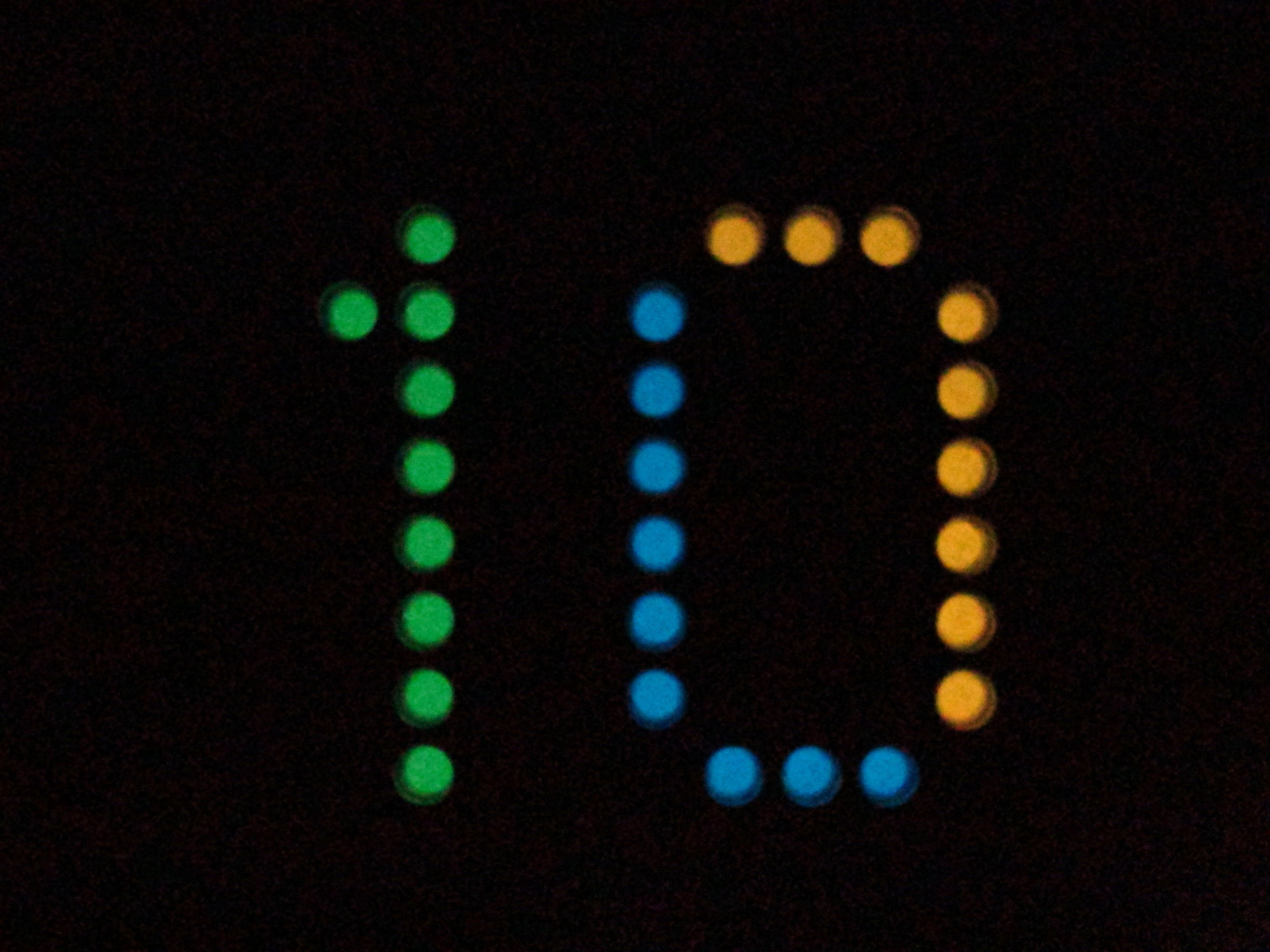
Akira Takai, Yasushi Okada, Masahiro Nakano and Takeharu Nagai, at Osaka University, used multicolour luminescent reporters to write the number 10, either by expressing them in bacterial cells streaked on an agar plate, or by aliquoting them in purified form in a 96-well plate.


10.2-Merge-v2-cropped2

{credit}Lauren Polstein and Charles Gersbach{/credit}

Lauren Polstein and Charles Gersbach at Duke University used light-sensitive transcriptional activators to photoactivate a GFP reporter in mammalian cells in the shape of the number 10.


Navneet Dogra and T. Kyle Vanderlick at Yale University examined bacteria stained with fluorescein (green) interacting with small unilamellar vesicles labeled in red. They used a laser to photobleach all fluorescence except that in the desired shape of the number 10. Image to come.

Finally, Kristina Woodruff and Sebastian Maerkl at EPFL used a standard microarrayer to spot live mammalian cells onto a 675-well array in the shape of a 10 (Woodruff et al, 2013).  Image to come.

We are very grateful to all contributors – thank you for helping us design a cover that salutes the creativity and ingenuity of methods developers!


Light sheet imaging in Nature Methods

It was only a few months before Nature Methods was launched in October 2004 that Jan Huisken and Ernst Stelzer had published a paper in Science in which they used light sheet microscopy – what they called selective plane illumination microscopy or SPIM – to image fluorescence within transgenic embryos. Simplistically put, this century-old technique achieves optical sectioning by illuminating a sample through its width with a thin sheet of light. In the last decade, Nature Methods has published a steady stream of papers reporting developments in light-sheet imaging. Here are the highlights.

Our very first light-sheet paper was also from the Stelzer group, reporting the use of deconvolution to improve resolution of the technique (Verveer et al, 2007). This was rapidly followed by a paper from Hans-Ulrich Dodt, in which samples such as entire insects or brain tissue were rendered transparent with clearing agents to produce spectacular light-sheet ‘ultramicrographs’ (Dodt et al, 2007). The push to higher resolution continued, with a paper from Albert Diaspro reporting 3D super-resolution imaging within thick samples using light-sheets (Cella Zanacchi et al, 2011). The Stelzer group, meanwhile, improved performance of the technique in larger samples that scatter more light, by combining it with structured illumination (Keller et al, 2010).

Thai Truong, Willy Supatto and Scott Fraser added two photon excitation to light-sheet imaging, thereby doubling the depth and increasing by an order of magnitude the speed at which they could image samples such as developing embryos with each approach alone (Truong et al, 2011); Supatto recently extended this to imaging in multiple colours  (Mahou et al, 2014). And then in 2012, the groups of Phillip Keller and Lars Hufnagel independently reported microscopes that could take take multiple views of a biological sample simultaneously, allowing rapid imaging of entire developing fly embryos at sub-cellular resolution (Tomer et al, 2012; Krzic et al, 2012).

Though light-sheet imaging is perhaps at its most powerful in the imaging of thick samples like embryos or tissue sections, it has been used for substantial performance improvements in cellular imaging as well. In 2011, Eric Betzig’s group used scanned Bessel beams to create thinner light sheets and thus much improved axial resolution, achieving isotropic 3D resolution and rapid imaging within living cells (Planchon et al, 2011). Note also that, as Tom Vettenburg, Kishan Dholakia and colleagues showed,  generating the light sheet using an Airy beam, rather than Gaussian or Bessel beam, yields an even larger field of view without sacrificing contrast and resolution (Vettenburg et al, 2014). Variations on the light-sheet theme have also been developed by the labs of Makio Tokunaga and Sunney Xie for single-molecule imaging within cells (Tokunaga et al, 2008Gebhardt et al, 2013).

In recent years, the excitement around this technology has been palpable, with several papers reporting impressive applications of light-sheet microscopy: it has been used to functionally image the entire fish brain (Ahrens et al, 2013) and the brain of ‘fictively behaving’ fish (Vladimirov et al, 2014), as well as to image the beating fish heart (Mickoleit et al, 2014).

Perhaps not surprisingly, the emphasis in methods development has also been shifting a little. On the one hand, platforms are being developed to make this valuable technique available more widely, for instance via the OpenSPIM or OpenSpinMicroscopy platforms (Pitrone et al, 2013; Gualda et al, 2013). At the same time, analytical tools are necessarily being developed to handle the vast reams of data that a light-sheet experiment generates. The group of Pavel Tomancak reported Bayesian-based deconvolution methods to analyse the large data sets that result from multiview imaging (Preibisch et al, 2014). Phillip Keller and colleagues described computational methods to segment and track nuclei in data sets from light sheet or other imaging, for fast lineaging of developing embryos (Amat et al, 2014). Misha Ahrens and colleagues reported Thunder, a suite of analytical tools built on a platform for distributed computing, enabling the mapping of brain activity in ‘fictively behaving’ zebrafish (Freeman et al, 2014).

It’s fair to say that this venerable method has been thoroughly revived over the past decade. Light-sheet imaging is poised to yield tremendous biological insight. We hope to keep you updated on future developments in Methagora.

Know your methods

In the September 2014 issue of Nature Methods, authors at the US National Institute of Standards and Technology argue in a Commentary that a productive way to frame the discussion about the reproducibility of biological results is to focus on how best to make good measurements. In other words, increasing the confidence in measurements is likely to also increase the reproducibility of the results of those measurements. Notably, in complex biological systems, making good measurements is not trivial. Read this month’s editorial introducing this topic here and link to the Commentary here.

Strengthening communities through competition

Community bioinformatics challenges help drive methods development.

Science moves ahead faster as a social enterprise, perhaps especially so in the dynamic area of bioinformatics. Bioinformatics competitions are important opportunities for developers (and users) to come together to define the essential questions in the field and decide on the best metrics to evaluate them. They also perform a critical function in making valuable benchmark datasets available to anyone, including small labs and young students.

At the end of a challenge, ideally, is a better appreciation of the most promising approaches to a problem, as well as a recognition of difficulties and opportunities for future development. And  there are new contacts for collaboration. As a reality check, it is less common for researchers with directly competing methods to collaborate; their work depends on a competitive funding model. But complementary approaches provide fertile ground for exploring new ways to attack a problem, and some contests are directly encouraging collaborative coding.

In our July Editorial, we continue our support of these initiatives, urging participation and an embrace of formats that maximize engagement among participants. Already, measures like on-line forums, webinars, and conferences involve participants in the planning and interpretation stages, which are critical for getting the most out of each event.

A variety of formats beyond the traditional bake-off are evolving in the collaborative spirit, encouraging more sharing of ideas and code. For example, hackathons take on more focused coding challenges in a single dedicated meet-up session, while open-source competitions make code available during the contest to allow researchers to learn from each other. These formats are not meant as an evaluation of existing methods, but promote new solutions. As Gustavo Stolovitzky of the DREAM challenges points out, publishing code during the event has the potential for ‘herding’ behavior (copy-the-leader), which can stifle creativity and produce a coding monoculture. A number of DREAM challenges now use a two-stage approach in which top performers from a traditional competition phase are invited back to develop a new and better solution together.

Journals and funders also play a role in supporting these efforts. Nature methods has published a number of papers resulting from community competitions (CAFA, DREAM, FlowCAP, Particle tracking and RGASP) and the Nature journals have been committed to providing these papers under a Creative Commons attribution-noncommercial-share alike unported license since January 2013.

There are difficulties associated with running large-scale events. Choice of data set and metrics can bias evaluations towards certain solutions, and the involvement of many developers can water down the conclusions resulting from the challenge. Moreover, usability is often not considered since it is hard to quantify. Ultimately, these issues can be helped by boosting participation in decision-making during planning stages, tailoring conclusions to each scenario that is tested, and having judging panels test the best-performing methods to ensure usability.

We are heartened to see the continued success of community-led competitions and the birth of contests in new areas. In a guest post, we invited organizers of the CAMI competition to announce their upcoming event on metagenome data interpretation.

Below, we provide a non-comprehensive list of some recent and ongoing challenges:

Bake-offs
Assemblathon – genome assembly
CAFA (Critical Assessment of Function Annotation) – protein functional prediction
CAGI (Critical Assessment of Genome Interpretation) – functional variant prediction
CAMI (Critical Assessment of Metagenome Interpretation) – see the announcement
CAPRI (Critical Assessment of PRediction of Interactions) – structure-based protein-protein interaction prediction
CASP (Critical Assessment of protein Structure Prediction) – protein structure prediction since 1994!
DREAM (Dialogue for Reverse Engineering Assessment and Methods) – systems biology challenges with hybrid formats and challenge-assisted review
FlowCAP (Flow Cytometry: Critical Assessment of Population Identification Methods)
Grand Challenges in biomedical image analysis
Particle tracking challenge
RGASP (RNA-seq Genome Annotation Assessment Project)

Crowdsourcing competitions, hackathons and fast challenges
BioHackathons – open-source programming meetups
Innocentive – commercial platform offering cash prizes (e.g. the $1 million US Defense Threat Reduction Agency (DTRA) challenge to identify organisms from a stream of DNA sequences)
DNA60IFX – short challenges based on DNA or RNA sequence data
DREAM – a number of recent and current challenges include a collaborative phase of tool development
Neurosynth hackathons – open-source programming meetups in computational neurobiology
Sequence Squeeze – open-source competition for sequence file compression (cash prize)
[topcoder] – variety of computational challenges, with some cash prizes

Sunset on the PSI

As discussed in this month’s Editorial, the Protein Structure Initiative (PSI), a 15-year, nearly $1 billion structural genomics project funded by the National Institute of General Medical Sciences (NIGMS), will be coming to an end in 2015. The impact of ending this project should be minimized to avoid the loss of valuable resources and expertise.

The PSI was begun in 2000 when the US NIH budget was in the midst of substantial growth, spurring the creation of many new “big science” projects. At that time, protein structure determination was painfully slow. The PSI’s initial goals were to develop tools and methods to improve the speed and ability to solve protein structures, as well as to generate a large resource of novel and unique protein structures to facilitate homology modeling and to promote follow-up functional studies.

Protein Structures

{credit}Image Credit: Erin Boyle{/credit}

The PSI drew criticisms from many in the structural biology community from its inception, however. Critics of the first two phases of the PSI pointed out that it focused mainly on solving small bacterial protein structures that were not biologically very interesting, simply because they were relatively easy to express, purify and crystallize. This led the PSI to substantially change course in the third and current phase, PSI-Biology, which began in 2010. The emphasis on throughput was diminished and shifted to solving important, difficult structures like human membrane proteins and drug targets. This change of course has led to several successful structures for highly interesting yet difficult proteins such as GPCRs.

In 2013, a scientific advisory panel produced a mid-point evaluation report of PSI-Biology for NIGMS (PDF), assessing its strengths and weaknesses. The panel commended the impressive number of high-quality structures and methodological advances of the PSI centers, but noted that outreach to the broader biological community was inadequate. The panel found that the PSI’s community resources – the Structural Biology Knowledgebase, a portal for research, news and resources produced by the PSI (in collaboration with Nature Publishing Group), and the Materials Repository, which provides over 80,000 plasmids and 106 empty vectors to the community – were not widely taken advantage of by researchers outside the PSI. The panel also noted that NIGMS should start planning to transition the PSI from its current set-aside funding structure to a different funding model that maintains its unique resources and capabilities – but that it should also extend PSI-Biology for another 3 to 5 years past 2015 to allow it to reach its full potential.

However, budget cuts and a reassessment of NIGMS’s large-scale research initiatives by its new director, Jon Lorsch, has led the institute to prematurely cut the program after the current phase, PSI-Biology, ends in 2015.

The loss of this large-scale program will certainly shake up structural biology in the US, particularly for all those involved in the PSI but also for the many researchers – even outside of traditional structural biology – who benefit from PSI resources. As we discuss in this month’s Editorial, NIGMS now has the opportunity to set an example for other funding agencies in how to wind down a big science project with minimal negative impact. Internal and external transition planning committees have been created by NIGMS to determine which resources and capabilities developed by the PSI should be preserved and how this can be done. NIGMS has also put out a Request for Information seeking community input on the utility of resources developed by the PSI; the response date (May 23, 2014) has only just passed.

NIGMS is expected to make a decision before the end of 2014 as to what will be done with the substantial high-throughput expression, purification and crystallization facilities developed during the PSI’s tenure. In the Editorial we argue that this infrastructure – and the large-scale raw data and metadata generated that is not in any database, but is valuable for mining and algorithm development – should be preserved as much as possible. We argue that a project to systematically sample protein folds should be continued on a smaller scale. We also argue that NIGMS should continue to facilitate team research – as the internal transition planning committee chair Douglas Sheeley has said it will – to tackle particularly challenging structural biology research problems that require hybrid methods to solve.

We will be watching closely to see whether the negative fallout from the end of the PSI can indeed be minimized.

Synthetic Biology at Nature Methods

Since its launch, Nature Methods has seen many papers that have influenced the Synthetic Biology community. As a supplement to our May Focus on Synthetic Biology we take a nostalgic trip through the highlights of our papers in this area for different aspects of synthetic biology.

Cloning
In 2007 Stephen Elledge and Mamie Li developed SLIC (sequence and ligation-independent cloning) a strategy that uses homologous recombination to assembly many DNA fragments in vitro in a single reaction. Later the same year Mitsuhiro Itaya and colleagues also used homologous recombination in their bottom up assembly to unite larger DNA pieces to genomes of ~ 140kb size.

In 2009 Daniel Gibson and colleagues presented their one-pot enzymatic reaction that successfully assembled genomes 100s of kilobases and has since been dubbed ‘Gibson Assembly’.  The method reached fame on Youtube when the Cambridge iGEM team for 2010 created a music video showing how Gibson Assembly saves frustrated scientists:

Gene and genome synthesis
In 2007, to improve error-free DNA synthesis, Duhee Bang and George Church developed circular assembly amplification that eliminated error-containing oligonucleotides from the assembly. A few years later  Jay Shendure and colleagues introduced their dial-out PCR to retrieve desired DNA molecules from a library  for gene assembly.

For an in depth review on the topic of DNA synthesis, error correction and gene assembly visit Sriram Kosuri and George Church’s review in our Focus issue.

In 2010, on the heels of their breakthrough with Mycoplasma mycoides JCVI-syn1.0 – the first chemically synthesized bacterial genome (Gibson D, et al Science 329, 2010) ­- Gibson et al. published the chemical synthesis of the mouse mitochondrial genome in our pages. They adapted Gibson Assembly to begin at the oligonucleotide level to rapidly make larger fragments that were then combined into the desired genome, exclusively in vitro.  Once synthesized a bacterial genome might need to be further modified, but to do so in an organism other than E. coli proved challenging. In 2013 Bogumil Karas et al, showed that whole genomes, as large as 1.8 megabases can be directly transferred from bacteria to yeast where genetic manipulation is routine.

In our current Focus issue Gibson reviews the state of the art in genome assembly techniques , compares strategies and discusses what the future may hold.

Genome modification
To quickly generate large libraries of promoters in targeted regions of a bacterial chromosome  George Church and colleagues presented coselection MAGE (multiplex automated genome engineering) in 2012.  The increasingly popular CRISPR system can also rapidly edit genomes with few off-target effects when Cas9 is used as a nickase as William Skarnes and colleagues showed earlier this year.

Gene activation can be tuned by targeting transcription factors via the CRISPR-Cas9 system as Charles Gersbach demonstrated in 2013.

Circuit design
To ease construction of complex circuits Adam Arkin and colleagues adapted known translational regulators to control transcriptional elongation in 2012.  A bit later the same year Jim Collins and colleagues showed that an iterative plug-and-play method makes use of a large repository of genetic components when designing circuits.  This year Jeff Tabor and colleagues showed how gene circuit dynamics can be controlled with light. On April 28 Douglas Densmore and his team introduced Raven , software that calculates assembly plans for complex circuits.

Parts characterization
To be successful in any of the above applications one needs reliable and well characterized parts. Last year Drew Endy, Adam Arkin and colleagues presented a method to quantify the performance of genetic elements and in a companion paper they introduced a library of standardized transcription and translation initiation elements available through biofab.

Towards the end of 2013 Christopher Voigt and colleagues expanded the designer’s toolbox with over 500 well characterized transcriptional terminators. Robert Landick discussed how these ‘better stop signs’ as he termed them provide insight into the mechanism of termination.

UPDATE: There is now a joint special on Synthetic Biology at nature.com/synbio with articles from Nature, Nature Reviews Microbiology and Nature Methods.

Enjoy reading. The papers mentioned above are listed below in chronological order.

Continue reading

What we publish

The editors of a scientific journal have an editorial prerogative to publish articles that fall under the editorial scope of the journal as they see it. But defining this scope in a way that is clear to those outside the editorial team can be difficult and any definition can become dated as science and the journal evolve. Here we discuss the scope of Nature Methods.

As stated in our Guide to Authors, Nature Methods publishes “novel methods and significant improvements to tried-and-tested basic research techniques in the life sciences.” We broadly define “research techniques” as methodological procedures, biological or synthetic reagents, computational algorithms, software tools, instrumentation and other technologies.

The phrase “basic research” in the sentence above is key and, as explained in April’s Editorial, methods intended for later stage research applied to the clinic, drug discovery or industrial processes are generally considered outside our scope. These applications are often classified as biotechnology and thus are probably more appropriate for Nature Biotechnology or, if clinically oriented, a Technical Report in Nature Medicine.

But as April’s Editorial acknowledges, method and tool developments can be relevant for both basic research and more ‘downstream’ applications. This requires us to be continuously walking an editorial tightrope between them. As circumstances change and fields develop we may need to adjust how we apply our editorial scope.

As also stated in our Guide to Authors, Nature Methods is targeted at “academic and industry researchers actively involved in laboratory practice.” The phrase “laboratory practice” is intended to indicate that the journal generally only publishes methods for work that occurs in a research laboratory environment. On occasion, we may consider compelling work that doesn’t fall under the typical definition of laboratory research, particularly in areas like ecology where the basic research environment extends beyond the confines of a brick and mortar lab. An example was our publication of The Metatron: an experimental system to study dispersal and metaecosystems for terrestrial organisms.

We are constantly reassessing our editorial scope and can work with authors to adapt the presentation of work that might otherwise be considered out of scope if we feel it is sufficiently compelling, relevant to our readership and can be appropriately presented as important for basic research. We are happy to respond to presubmission inquiries submitted via our manuscript submission system asking about the appropriateness of the scope of a proposed manuscript. But if a manuscript is already written please submit the full manuscript as a regular submission and don’t worry about formatting it to fit Nature Methods’ article style. This will allow us to make a more informed decision and format can be dealt with later in the event we proceed towards publication. If a manuscript is clearly out of scope we will endeavor to provide a fast decision.

Finally, if there is an area of basic biological research that you feel is underrepresented in Nature Methods but should be of substantial interest to our readership, please let us know. For example, we published virtually no computational methods for the first several years of the journal but they now represent a substantial fraction of our articles. As we strive to serve our readers we want to avoid falling into a pattern of publishing research limited to a few areas, but our success in doing this depends heavily on receiving submissions from a broad range of research areas and we encourage the wider basic research community to consider Nature Methods even if we haven’t yet published much, or anything in a particular area.

Here there be software

Software plays an important role in scientific research, and published studies increasingly rely on custom software code developed by authors. This calls for better transparency in research articles and improved access to the software and code itself.

This month in Nature Methods and on methagora we revisit issues regarding software reporting and availability first raised exactly seven years ago in our March 2007 Editorial “Social software“. Our March 2014 Editorial updates and expands on these editorial policies and a blog post provides details of our guidelines for custom algorithms and software reported in Nature Methods research papers. We encourage researchers to read these, particularly those considering submitting a research manuscript using or reporting custom software to us. We also hope that publicizing our editorial policies might aid other journals in thinking about how to handle algorithms and software associated with research they publish.

Of course, these efforts are only one small part of what needs to be done to improve access to and use of scientific research software. As can be seen by our somewhat complex guidelines, it is difficult to establish simple rules that are sensible and fair for all cases and all communities. Community participation will be essential for refining and improving how software is handled.

Nature Methods currently relies on the use of Supplementary Software zip files for authors to supply the software and code underlying research articles. This isn’t pretty but it fulfills our basic needs. For example, 50% of the research articles in our March issue contain Supplementary Software files. But better methods are needed to archive and document code and assign provenance.

An important initiative in this regard is the “Code as a research object” project that is a collaboration between Mozilla Science Lab, Github and figshare that seeks to “better integrate code and scientific software into the scholarly workflow.” The aim is to create citable endpoints for the exact code used in particular studies. [Full disclosure: figshare is a product of Digital Science which, like Nature Methods, is part of Macmillan Publishers.]

The project is still in its early stages and follows on the similar but broader Research Object community project. Similarly, GigaScience and F1000Research are experimenting with archiving code and pipelines with DOIs.

We applaud these efforts and encourage the broader research community to participate in them. The current discussion about what is needed for code reuse (announced on the ScienceLab blog) and going on in a thread at Github would greatly benefit from more input by researchers who don’t consider themselves code jockeys.

There are many sophisticated and powerful things that could be done in an ideal world to facilitate code exposure and reuse, but the situation at the great majority of journals is so underdeveloped and the needs so acute that even small flexible steps forward will have a positive impact. Most important is for facilities to be put in place that allow and encourage the entire community to move forward, not just a small portion of it.

Guidelines for algorithms and software in Nature Methods

A large proportion of original research published in Nature Methods relies to varying degress on custom algorithms and software developed by the authors. Here we provide guidance on our relevant material sharing and reporting policies.

Nature Methods first outlined our material sharing and reporting standards for algorithms and software in a March 2007 Editorial. Now, after seven years of experience applying those policies we updated and expanded on them in our March 2014 Editorial. On this page we provide more detailed guidelines for authors submitting manuscripts containing unpublished algorithms and software they created. We are posting this information here because we’d like these guidelines to evolve and we want input from our communities on how they think this should happen. Please comment below and let us know your thoughts. We will update this document as our policies change.

Manuscripts published in Nature Methods include methods and tools in which algorithms and software represent an increasingly important methodological component. However, the degree to which they are central to the reported methodology can vary considerably. The algorithm or tool may be the entire motivation for publishing the work or it may be ancillary to it. Additionally, the methodology may be a novel algorithm of value in and of itself but a coded implementation is still necessary for the authors to show that it works as expected. Finally, the software tool may implement existing algorithms in a user-friendly form to deliver high value functionality of substantial general interest. Because of this wide variety it is inappropriate to enforce one-size-fits-all standards for algorithms and software reported in Nature Methods. The guidelines below represent our current editorial position on software reporting and release.

Client-side Software
This is software that is installed and used on a personal computer and not intended to be accessed remotely as a web service. It can be entirely stand-alone on a commonly available operating system (Windows, Mac OS X, or *nix) or can require the user to have a popular software platform installed (MATLAB or LabVIEW). In all cases, but particularly when using MATLAB or LabVIEW, all platform versions and software dependencies must be detailed in the supplied documentation.

At Submission

  • If the custom algorithm/software is central to the method and has not been reported previously in a published research paper it must be supplied by the authors in a usable form including one or more of the following.
    1. Source code
    2. Complete pseudocode
    3. Full mathematical description of the algorithm
    4. Compiled standalone software

    We strongly urge that full source code be provided. A compiled executable alone is not sufficient but may be required if the tool is intended to be of wide general use. Final acceptable forms of release of the algorithm, software and code will be determined by the editor after consultation with referees. This decision will be influenced by the editorial motivation for publishing the work (i.e. high novelty, satisfies wide general need, etc).

  • If the software is ancillary to the methodology being reported or is a routine implementation of obvious processes, such as microscope control software or analyses that are otherwise adequately described, the software need not be supplied to reviewers at submission but final release requirements may change in the course of the review process.
  • Supplied source code or software must be accompanied by documentation sufficient for a typical user to compile, install and use the software. Depending on the nature of the software tool, how central it is to the manuscript and our editorial motivation for considering the work, the minimum documentation may be a simple readme file or a full manual in PDF format.
  • If appropriate, sample data known to work on the software should be provided along with the expected output. Referees are encouraged to try and use the tool to analyze their own data.
  • The software and associated files may be supplied for reviewers as either:
    1. A single Supplementary Software zip file up to 200 MB in size
    2. Four DVDs to be mailed to the reviewers.
  • Any restrictions on the availability of software or code used to implement novel algorithms must be specified at the time of submission. Editors will decide whether any restrictions are acceptable in consultation with the reviewers. If some restrictions are deemed acceptable, they must be clearly explained in the methods section of the manuscript. Authors must supply all information needed for the reviewers to properly evaluate the software or code. If the motivation of the submitted manuscript is to provide a useful tool, rather than report a new algorithmic development, there should be no substantial restrictions on software or code availability.
  • We encourage authors to provide a license with the software or code.
  • A narrative description of key algorithmic components should be provided in the main text. Extensive equations, pseudocode or snippets of source code should be confined to the Online Methods or a Supplementary Note.

At Acceptance

  • If the software is central to the methodology and non-obvious, the source code should be provided in a Supplementary Software zip file as described above so that readers can easily access the exact code used to obtain the results in the paper. There are some possible exceptions:
    1. If the author’s institution requires a user to accept a license agreement or if the author has other reasonable grounds for not providing the source code as Supplementary Software, it may be acceptable for the author to host source code on an institutional server and require that users fill out an online form and agree to a license before downloading the software. In this instance the software must have version numbering and a link to the version used in the work must be provided in the manuscript.
    2. In some situations it may be permissible for authors to supply only compiled software as Supplementary Software but the source code to academic users upon email request. Details of availability must be clearly stated in the manuscript.
    3. It is not acceptable to make software and code available by email request only.

  • If the software or code isn’t the main tool/method being reported in the manuscript the authors may provide a note in the readme file of the Supplementary Software cautioning users that the code is unsupported and not intended for general use. In this case it is permissible that the software or code be made available only by email request but the authors must state this availability in the manuscript.
  • Regardless of how the software is made available, the code supplied with the manuscript must be identical to that used to obtain the data in the paper. An exception can be made for changes that don’t alter the processing of input data. The authors may however provide a link to access new versions of the software.
  • We strongly encourage authors to include a license with all published software and code.
  • We encourage authors to provide macros for recording the software version and parameter settings during analyses or to integrate this functionality into the software itself.

Web Tools/Resources
These represent a special class of software that many times can’t be expected to follow the same guidelines outlined above. This is particularly true if the web tool or resource is being supplied as a service and has few, if any, novel computational aspects to it. The only end-user requirement for web tools is that they be freely accessible with any modern web browser.

Nature.com provides a proxy server for reviewers to access web tools and resources anonymously.

At Submission

  • The authors must supply a working link and any necessary log in information.
  • Any unpublished algorithms central to the operation of the tool should be supplied in forms a), c) or d) detailed above.

At Acceptance

  • The authors should supply written confirmation that they will keep the website and tool operating and freely accessible for the foreseeable future.

Is phototoxicity compromising experimental results?

Light-induced damage to biological samples during fluorescence imaging is known to occur but receives too little attention by researchers.

The December Technology Feature in Nature Methods asks if super-resolution microscopy is right for you and a point that comes up repeatedly from the researchers we interviewed is the danger of phototoxicity and photodamage caused by the high irradiation intensities needed for the illuminating light. This has long been a concern with these methods and many of the papers describing them mention it.

But as discussed in the December Editorial, even fluorescence microscopy with low irradiation intensities can cause dangerous levels of phototoxicity that permanently damage the sample. Microscopists are aware of these concerns but there has been little effort to implement processes intended to reduce the likelihood of it compromising research study results. Dave Piston, Director of the Biophotonics Institute at Vanderbilt University School of Medicine, laments that while phototoxicity is a big deal he has gotten zero traction with NIH reviewers on trying to build some rules for it.

There are some good resources available to researchers that highlight the dangers of phototoxicity and provide advice on how to limit it. Methods in Cell Biology Vol 114 has an excellent chapter by Magidson and Khodjakov, Circumventing Photodamage in Live-Cell Microscopy, that should be mandatory reading for all researchers using fluorescence microscopy for biological research. Also, Nikon’s MicroscopyU has a literature list with several dozen references and recommended reading on phototoxicity. It could use some updating but is still useful.

Despite the amount of microscopy literature that discusses phototoxicity, discussion of the phenomenon in research articles published in Nature Journals is conspicuously absent. This is highlighted by a simple full-text search we performed on the HTML versions of research articles published in Nature, Nature Cell Biology, Nature Immunology, Nature Methods and Nature Neuroscience. The articles retrieved were limited to original research articles.

The table below lists the number of occurrences of each of the listed words in the period from January 1, 2005 to November 3, 2013 in each of the indicated journals. The percentages represent the number fraction of articles containing ‘phototoxicity’ relative to the numbers of articles containing each of the microscopy- or fluorescence-related terms. Note that this is NOT a measure of co-occurrence, only a measure of how common the term ‘phototoxicity’ is relative to the other terms.

phototoxicity fluorescence fluorescent microscopy microscope
# # % # % # % # %
Nature 8 2120 0.4% 1925 0.4% 1995 0.4% 1918 0.4%
Nature Cell Biology 8 815 1.0% 728 1.1% 866 0.9% 822 1.0%
Nature Immunology 6 552 1.1% 574 1.0% 408 1.5% 326 1.8%
Nature Methods 27 565 4.8% 494 5.5% 441 6.1% 407 6.6%
Nature Neuroscience 18 639 2.8% 727 2.5% 587 3.1% 736 2.4%


The same analysis was repeated with the term ‘photodamage’ to determine if there was a substantial difference in the usage of these two similar terms.

photodamage fluorescence fluorescent microscopy microscope
# # % # % # % # %
Nature 18 2120 0.8% 1925 0.9% 1995 0.9% 1918 0.9%
Nature Cell Biology 6 815 0.7% 728 0.8% 866 0.7% 822 0.7%
Nature Immunology 2 552 0.4% 574 0.3% 408 0.5% 326 0.6%
Nature Methods 29 565 5.1% 494 5.9% 441 6.6% 407 7.1%
Nature Neuroscience 12 639 1.9% 727 1.7% 587 2.0% 736 1.6%


These results carry the potentially large caveat that the analysis did not include the text of the supplementary information, but the rarity with which phototoxicity or photodamage is discussed (0.4% to 7% relative to microscopy terms) suggests that researchers don’t appreciate how important it is to pay attention to artifacts that result from light irradiation. Luckily, there are exceptions to this state of affairs.

An excellent example of testing for phototoxicity and the subtle effects it can induce can be found in a manuscript from Jeff Magee’s lab at Janelia Farm Research Campus published last year in Nature. Quoting from the manuscript, “Particular care was taken to limit photodamage during imaging and uncaging. This included the use of a passive 8× pulse splitter in the uncaging path in most experiments to reduce photodamage drastically [Ji, N. et al. Nat. Methods (2008)]. Basal fluorescence of both channels was continuously monitored as an immediate indicator of damage to cellular structures. Subtle signs of damage included decreases in or loss of phasic Ca2+ signals in spine heads in response to either uncaging or current injection, small but persistent depolarization following uncaging, and changes in the kinetics of voltage responses to uncaging or current injection. Experiments were terminated if neurons exhibited any of these phenomena.”

It is easy to see how these changes in Ca2+ responses could easily have been interpreted as real biological effects caused by the uncaged glutamate, rather than the uncaging light itself.

It is unrealistic to expect that any mandates or oversight would be able to prevent or detect such consequences of phototoxicity in research studies. It is essential that investigators themselves be vigilant and implement appropriate controls to detect these effects. Na Ji, also at Janelia Farm Research Campus says, “It is not enough to only look for instant and dramatic signs of phototoxicity. Sometimes the effects may be more subtle and even unperceivable during the imaging period, but may become obvious when the same sample is imaged the next day. Care has to be taken in data collection and interpretation, especially when the biological process under investigation itself is a subtle one.”

Finally, the application is just as important as the imaging method being used. For example, light-sheet microscopy is excellent at reducing irradiation levels in volumetric imaging. But some applications of super-resolution microscopy, even on living samples, might be less susceptible to artifacts caused by phototoxicity than are sensitive long-term imaging applications of living samples by light-sheet microscopy. Nobody’s microscope earns them a free pass on the dangers of photodamage arising from phototoxicity. Everyone needs to be vigilant.

Update: A reader helpfully pointed out that the danger of phototoxicity and photodamage also applies to optogenetics, where light (often in the blue region of the spectrum) is used to control protein activity.