Positive feedback drives network (and manuscript) maturation.

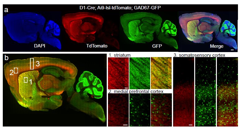
Whole-brain anatomical mapping of D1-Cre expression in inhibitory neurons (from Supp Fig.2)

It really is an embarrassment of riches here at Nature these days, what with so many excellent neuroscience-related studies emerging. Just in the last couple of weeks, we’ve had the following studies:

So really, a lot to write about from a science perspective. However, this blog is dedicated to bringing you the editorial back-story, so I wanted to touch on yet another interesting study, published in print today. This new paper offers an opportunity to discuss an important editorial issue: the manuscript appeal process. For more details, you can always read the appropriate section in our guide to authors. But it’s often helpful to follow a particular [successful] example in order to illustrate the process. Continue reading

“There is no spoon…”: Paralyzed fish navigates virtual environment while we watch its brain

Overlaid on the micrograph of the fish is a slice of its brain measured with a laser scanning microscope, in which single neurons are visible.{credit}(courtesy of Ahrens et al.){/credit}

Sometimes an experiment will just reach off the page and slap you in the face, demanding attention. This happens to me every so often and I must admit, our latest paper from the lab of Florien Engert induced such an experience. There have been several cool, technical tours-de-force (is that proper grammar??) over the last few years involving different creatures navigating in a virtual environment while neuronal activity was monitored. These include a mouse running on a spherical treadmill, as well as a fly marching along a similar treadmill-style ball. But in these examples, having the subject head-fixed (for the stability of recordings in the brain, either with electrodes or through imaging) was moderately non-intrusive since walking motions were independent of the head. The same can’t be said for the subject in this latest example of a virtual reality navigator: a wriggling, swimming fish. Therefore, a more creative solution had to be sought and in a paper published online yesterday, Ahrens, Engert and colleagues decided that paralysis was the way to go in order to follow the neural activity of this navigating fish. Continue reading

Parietal decision sequences – and more of mice and monkeys

From Figure 2 of Harvey et al

Back in the 1990’s, one of the most intense battlegrounds in systems neuroscience was in monkey posterior parietal cortex. Labs competed to claim what a little strip of cortex called lateral intraparietal area (LIP) really does – decision, movement planning, attention, reward, or all of the above – mostly using single cell recording in behaving monkey. The experiments were (and still are) tough: standard operating procedure requires a well-trained monkey who will perform hundreds if not thousands of trials a day and then isolating neurons one at a time to find ones that respond during some interesting part of the trial. And then lots and lots of repetition so that you can average over many neurons. All things considered, it’s remarkable how much the field has been able to learn with this toolbox.

Fast forward to present day, there’s a new kid on the block. As I discussed a few weeks ago, rodent behavior and physiology is booming. People are taking on questions previously studied mainly in primates and are taking full advantage of the recent storm of new techniques. This is typified by today’s paper by Chris Harvey, Philip Coen, and David Tank, which goes back to the question – what does posterior parietal cortex do during a decision task?  They imaged populations of neurons while mice used visual cues to navigate a virtual maze. Just like in primates, individual neurons were selective for different choices that the mouse made. But unlike in primate parietal cortex, where neurons tend to have sustained responses leading up to the time of decision, individual neurons responded transiently in different portions of the trial. So as a population, different choices were represented by distinct sequences of neuronal activity. This kind of sequential firing has been seen in other parts of the rodent brain such as hippocampus, but not in posterior parietal cortex.  Continue reading

The Fine Architecture of Learning and Joint Publication

(image courtesy of Svoboda lab, https://openwiki.janelia.org/wiki/display/SvobodaLab/Research)

You warily walk into a dark compartment, wondering if there is food inside. Suddenly there is a loud tone and you feel an uncomfortable surge of electricity through your feet. This goes without saying, but it won’t take long before you will learn to be afraid of that tone. However, over time, you hear the tone without the shock, and slowly (foolishly??) accept that the previous connection may no longer hold.

Or perhaps you are extremely motivated to work for food, given that in your home area, nutrition has been sparse and hard to come by. You see millet seeds seemingly just within the reach of your fore-limb. Though not a typical movement for you, you reach for it. In another instance, you find a different type of food that is difficult to handle. However, it is nourishment nonetheless, so you will learn the required motor skills.

SPOILER ALERT: In each of the above cases, you were a mouse the whole time (I know!) But this is a neuroscience blog, not M.Night Shyamalan’s IMDB page, so perhaps we should focus on what was taking place in the brain as each scenario played out. In both of the cases above, learning was occurring, with new information stored away within the appropriate neural connections of particular brain areas. These situations are on display in a pair of new(ish) papers out in Nature, exploring the structural substrates of such learning and identifying patterns underlying the observed structural changes as learning occurred. Continue reading

Layer magic and monkey business

Layers of human cortex drawn by Ramon y Cajal. Image from Wikimedia Commons

We’ve known for over a century that sensory cortex is arranged in distinct layers, each containing a different make up of neuronal types and projection patterns, but we don’t actually know that much about the actual computations performed in each layer.  Today a paper from Massimo Scanziani’s lab takes a big step towards cracking the function of the bottom layer (layer 6) in mice. Layer 6 neurons project both to upper cortical layers and to the lateral geniculate nucleus in the thalamus, which itself is the primary input to cortex, and so are primed to play a large modulatory role. Using a monumental combination of optogenetics, intracellular recording, and behavioral testing, the paper convincingly makes the case that layer 6 controls the gain of visual responses of upper layer neurons (i.e. changes the size of their responses without altering their selectivity). Gain control is a fundamental computation in cortex, and has been invoked as a mechanism for attention, perception, spatial processing, and more. The cellular mechanism here is worked out in primary visual cortex, but it could potentially operate throughout layered cortex.

Continue reading