Optogenetics in neuroscience at Nature Methods

The optogenetic manipulation of cellular properties has not only revolutionized neuroscience, but this technology can also be applied to the manipulation of signaling pathways, transcription or other processes in non-neuronal cells. Here, we highlight some of the papers we have published on the neuroscience side of optogenetics.

Optogenetic tools

2014 has been an exciting year for us with the publication of new optogenetic tools. Klapoetke and Boyden developed Chrimson and Chronos, two channelrhodopsins that they discovered in a screen of algal transcriptomes. Chrimson is more red-shifted than previously known channelrhodopsins while Chronos has faster kinetics. Hochbaum and Cohen described another algal channelrhodopsin called CheRiff, which is highly sensitive to blue light stimulation, making it compatible with red-shifted voltage sensors.

Previously, we published papers describing modifications to optogenetic tools. For example, Prakash and Deisseroth tailored opsin with custom properties. To ensure stoichiometric expression of optogenetic activators and/or inhibitors, Kleinlogel and Bamberg simply and elegantly fused the two proteins into a single chain. Depending on the two partners, this marriage can lead to synergisms or bidirectional effects. Finally, Mattis and Deisseroth undertook a comprehensive characterization of available tools.

Optogenetic applications

Since the initial description of Channelrhodopsin2 (ChR2) as an efficient tool to evoke neural activity in a light-dependent manner, we have seen a flurry of papers applying ChR2 for a variety of questions in neuroscience. For instance, Zhang and Oertner combined this tool with two-photon calcium imaging in rat slices to study synaptic plasticity. Liewald and Gottschalk applied the same methodology to analyze synaptic function in freely moving C. elegans.

ChR2 can also be used to map the function of brains regions as Ayling and Murphy demonstrated by evoking activity in limb muscles via light stimulation in the motor cortex of ChR2 transgenic mice. Similarly, Guo and Ramanathan mapped neural circuitry in C. elegans by combining ChR2-mediated neural activation with imaging of a genetically encoded calcium sensor in downstream neurons. To facilitate circuit mapping in mice, Zhao and Feng generated mouse lines that express ChR2 in GABAergic, cholinergic, serotonergic or parvalbumin-expressing neurons.

While ChR2 is a very popular tool in optogenetics, other family members can do the job as well. C1V1T is a fusion of two different opsins and is particularly useful when applying two-photon excitation, as shown by Packer and Yuste. ReaChR is activated by red light and thus especially useful in vivo. Inagaki and Anderson studied courtship behavior in Drosophila with this tool.

Method of the Year

We celebrated the impact of optogenetics by recognizing the technology as our Method of the Year 2010. We marked the occasion with the publication of special Commentaries on the subjects. Deisseroth discussed the past, present and future of optogenetics. Hegemann and Möglich deliberate on the exploration of new optogenetic tools. And Peron and Svoboda illuminated us on the precise delivery of optogenetic stimulation. In addition, our News Feature recounted the stories behind the “Light tools”.

If we have sparked your interest, the mentioned papers are listed below.

We are excited to hear about the upcoming developments in optogenetics from you.


Nathan C Klapoetke, Yasunobu Murata, Sung Soo Kim, Stefan R Pulver, Amanda Birdsey-Benson, Yong Ku Cho, Tania K Morimoto, Amy S Chuong, Eric J Carpenter, Zhijian Tian, Jun Wang, Yinlong Xie, Zhixiang Yan, Yong Zhang, Brian Y Chow, Barbara Surek, Michael Melkonian, Vivek Jayaraman, Martha Constantine-Paton, Gane Ka-Shu Wong & Edward S Boyden
Independent optical excitation of distinct neural populations
Nature Methods 11, 338–346 (2014) doi:10.1038/nmeth.2836

Daniel R Hochbaum, Yongxin Zhao, Samouil L Farhi, Nathan Klapoetke, Christopher A Werley, Vikrant Kapoor, Peng Zou, Joel M Kralj, Dougal Maclaurin, Niklas Smedemark-Margulies, Jessica L Saulnier, Gabriella L Boulting, Christoph Straub, Yong Ku Cho, Michael Melkonian, Gane Ka-Shu Wong, D Jed Harrison, Venkatesh N Murthy, Bernardo L Sabatini, Edward S Boyden, Robert E Campbell & Adam E Cohen
All-optical electrophysiology in mammalian neurons using engineered microbial rhodopsins
Nature Methods 11, 825–833 (2014) doi:10.1038/nmeth.3000

Rohit Prakash, Ofer Yizhar, Benjamin Grewe, Charu Ramakrishnan, Nancy Wang, Inbal Goshen, Adam M Packer, Darcy S Peterka, Rafael Yuste, Mark J Schnitzer & Karl Deisseroth
Two-photon optogenetic toolbox for fast inhibition, excitation and bistable modulation
Nature Methods 9, 1171–1179 (2012) doi:10.1038/nmeth.2215

Sonja Kleinlogel, Ulrich Terpitz, Barbara Legrum, Deniz Gökbuget, Edward S Boyden, Christian Bamann, Phillip G Wood & Ernst Bamberg
A gene-fusion strategy for stoichiometric and co-localized expression of light-gated membrane proteins
Nature Methods 8, 1083–1088 (2011) doi:10.1038/nmeth.1766

Joanna Mattis, Kay M Tye, Emily A Ferenczi, Charu Ramakrishnan, Daniel J O’Shea, Rohit Prakash, Lisa A Gunaydin, Minsuk Hyun, Lief E Fenno, Viviana Gradinaru, Ofer Yizhar & Karl Deisseroth
Principles for applying optogenetic tools derived from direct comparative analysis of microbial opsins
Nature Methods 9, 159–172 (2012) doi:10.1038/nmeth.1808

Yan-Ping Zhang & Thomas G Oertner
Optical induction of synaptic plasticity using a light-sensitive channel
Nature Methods 4, 139 – 141 (2006) doi:10.1038/nmeth988

Jana F Liewald, Martin Brauner, Greg J Stephens, Magali Bouhours, Christian Schultheis, Mei Zhen & Alexander Gottschalk
Optogenetic analysis of synaptic function
Nature Methods 5, 895 – 902 (2008) doi:10.1038/nmeth.1252

Oliver G S Ayling, Thomas C Harrison, Jamie D Boyd, Alexander Goroshkov & Timothy H Murphy
Automated light-based mapping of motor cortex by photoactivation of channelrhodopsin-2 transgenic mice
Nature Methods 6, 219 – 224 (2009) doi:10.1038/nmeth.1303

Zengcai V Guo, Anne C Hart & Sharad Ramanathan
Optical interrogation of neural circuits in Caenorhabditis elegans
Nature Methods 6, 891 – 896 (2009) doi:10.1038/nmeth.1397

Shengli Zhao, Jonathan T Ting, Hisham E Atallah, Li Qiu, Jie Tan, Bernd Gloss, George J Augustine, Karl Deisseroth, Minmin Luo, Ann M Graybiel & Guoping Feng
Cell type–specific channelrhodopsin-2 transgenic mice for optogenetic dissection of neural circuitry function
Nature Methods 8, 745-752 (2011) doi:10.1038/nmeth.1668

Adam M Packer, Darcy S Peterka, Jan J Hirtz, Rohit Prakash, Karl Deisseroth & Rafael Yuste
Two-photon optogenetics of dendritic spines and neural circuits
Nature Methods 9, 1202–1205 (2012) doi:10.1038/nmeth.2249

Hidehiko K Inagaki, Yonil Jung, Eric D Hoopfer, Allan M Wong, Neeli Mishra, John Y Lin, Roger Y Tsien & David J Anderson
Optogenetic control of Drosophila using a red-shifted channelrhodopsin reveals experience-dependent influences on courtship
Nature Methods 11, 325–332 (2014) doi:10.1038/nmeth.2765

Karl Deisseroth
Optogenetics
Nature Methods 8, 26–29 (2011) doi:10.1038/nmeth.f.324

Peter Hegemann & Andreas Möglich
Channelrhodopsin engineering and exploration of new optogenetic tools
Nature Methods 8, 39–42 (2011) doi:10.1038/nmeth.f.327

Simon Peron & Karel Svoboda
From cudgel to scalpel: toward precise neural control with optogenetics
Nature Methods 8, 30–34 (2011) doi:10.1038/nmeth.f.325

Monya Baker
Light tools
Nature Methods 8, 19–22 (2011) doi:10.1038/nmeth.f.322
Uncertain of the future, three ALS patients spearhead a new fund

It was only last summer, while on a kite surfing holiday, Garmt van Soest observed that his right hand was unusually weak. He also noticed that his speech was gradually becoming slower. “You wouldn’t know it now but I was really the fastest speaker in the office,” he says, enunciating deliberately. The changes motivated him to see his doctor. “I was really lucky,” says van Soest, a senior manager in Accenture Strategy based in Amsterdam. “I was diagnosed with ALS [amyotrophic lateral sclerosis] in six weeks. For most patients, the process takes a year.”

Since his diagnosis in August of 2013, van Soest has been using his management consulting background to strategize how best to contribute to the ALS community. He soon met two fellow ALS patients and entrepreneurs, Robbert Jan Stuit and Bernard Muller. On 19 May the three launched an ALS-specific investment fund, called Qurit Alliance. Qurit Alliance aims to raise €100 million ($139 million) to then invest into ALS-focused private biotechnology companies and institutions to kick start projects of drug discovery and smarter design drug trials to find ALS treatments.

“This is one of the novel, innovative ventures that wants to make sure orphan disease clinical pipelines do not dry up as the pharma model and venture investment shifts to later stage opportunities,” says Steve Perrin, CEO of the Massachusetts-based ALS Therapy Development Institute.

Continue reading

Naked Neuroscience: Dr Hannah Critchlow reflects on educating the public about Neuroscience

Hannah Critchlow: Educating people across the globe on the values of neuroscience.

Hannah Critchlow: Educating people across the globe on the values of neuroscience.

Dr Hannah Critchlow is a neuroscientist with a background in neuropsychiatry. She currently strips down the brain with the BBC broadcast Naked Scientists. Using radio, on-line channels and live events she designs, produces and presents a neuroscience-focused, interactive multimedia experience for the public.

In 2014 Hannah was named as a ‘Top 100 UK scientist’ by the Science Council for her work in science communication. In 2013 she was named as one of Cambridge University’s most ‘inspirational and successful women in science’. During her PhD she was awarded a Magdalene College, Cambridge University Fellowship, and as an undergraduate received three University prizes as Best Biologist. She previously worked as Strategic Manager for Cambridge Neuroscience and on secondment with the British Neuroscience Association.

Hannah’s choice of career stemmed from working as a nursing assistant at St Andrews Psychiatric Hospital. When not being enthused by all things brainy, Hannah spends her time splashing about by the river and living the houseboat dream.

For neuroscientist Dr Hannah Critchlow, the last 12 months have been pretty special. If being named one of Cambridge University’s most ‘inspirational and successful women in science’ late in 2013 wasn’t surprise enough, what was to follow was in her words, even more “gobsmacking.” Just days later, while in New Zealand for Christmas, Dr Critchlow was recognised by the Science Council as one of the top 100 UK scientists for her work in science communication.

Continue reading

Brain initiatives galore, smiles aplenty

Vivien Marx reports on the Society for Neuroscience meeting in San Diego and the big brain projects in the EU and US.

SfN attendance sign

The Society for Neuroscience annual meeting in San Diego clocked record attendance.{credit}Vivien Marx{/credit}

The brain is hot.

Despite dismay about the recent 16-day US government shutdown, the impact of automatic budget cuts–the sequester–taking effect in light of federal budget disagreements in Washington, and the general economic malaise, there is palpable excitement. New large-scale initiatives are getting underway around the world to develop technologies to empower neuroscientists.

This year’s Society for Neuroscience (SfN) meeting in San Diego that has just ended, clocked a record attendance of over 30,000 attendees, noted society president Larry Swanson to attendees with a broad smile in one of his conference announcements. “It is an inspirational time to be a neuroscientist,” he said, with the field drawing attention, for example, across the European Union and in the White House. In a town hall meeting for the Brain Research through Advancing Innovative Neurotechnologies (BRAIN) Initiative, there was no lack of critical comments and suggestions of aspects to include in BRAIN. But smiles stayed plentiful as funders explained their plans.

The fact that the US president chose neuroscience as his multi-year, signature project is something “we should all be pretty excited about,” says Tom Insel, director of the National Institutes of Mental Health. In addition to projects in the US, such as  (BRAIN) Initiative and the EU’s Human Brain Project, large neuroscience projects are just emerging in Australia, China, Japan and Israel. “This is beginning to feel like a global movement,” he says. And projects are unfurling in the private sector, too.

The new tools, says Story Landis, director of the National Institute of Neurological Disorders and Stroke, will help neuroscientists do their work “bigger, better, faster” and expand the research strides made in recent years.

Much remains to be done. Compared to what is known about the kidney or heart, very little is known about the brain, says Insel. Adding to the neurological diseases, he noted, are the “invisible wounds of war” such as traumatic brain injury and post-traumatic stress disorder. Tools to help diagnose these illnesses are urgently needed.

Nora Volkow, director of the National Institute of Drug Abuse says that the BRAIN initiative stands to “act like a catalyst” in ways not unlike the decoding of the human genome and its successive “avalanche of discovery.”

Besides attending SfN’s hundreds of sessions and 17,000 posters, scientists had the chance to get up close and personal with representatives from the funding agencies and to hear about and discuss the new opportunities. Here is a snapshot of some of the announcements.

European Union
As Daniel Pasini from the European Commission’s programme on future and emerging technologies explained, the 10-year European Human Brain Project has invited the scientific community to present “grand ideas” for a massive effort to computationally reconstruct the human brain using supercomputers.

The model will help to study brain-related diseases, which are a major health challenge, an economic and social burden, and to pool data and expertise more effectively and translate results for treatments.

The project, which took three years of planning, involves over 250 scientists across Europe in 135 research groups in 22 countries, including groups in the US and Asia. The program began officially in October and has a budget of $1.6 billion. Half of the money will come from the EU the other will come from national funding sources, Pasini says. The first phase is slated to last 30 months and is funded with $100 million.

Six platforms are to be developed including, for example, the neuroinformatic platform as a single point of access to all neuroscience and clinical data along with software tools. The other platforms involve brain simulation, high performance computing, medical informatics, neuromorphic projects and neurorobotics. The idea is to keep improving the model as new data become available. All tools and data are set to be made available to the global scientific community. The plan is to create the ‘CERN for brain research.’ Not unlike a telescope facility or a super-collider, scientists will be able to perform experiments and use this platform to help continue to expand the model.

Deconstructing Henry

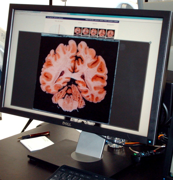
The Brain Observatory at UC San Diego is running ‘Deconstructing Henry’ an examination of the Brain of patient H.M.{credit}Vivien Marx{/credit}

US Defense Advanced Research Projects Agency (DARPA)
“Yes, we build guns and bombs, that is true,” says Colonel Geoffrey Ling of DARPA more generally. He is a neurologist who also served in Afghanistan and Iraq and currently deputy director of DARPA’s division responsible for defense sciences, which does not build bombs and guns. He and many other neuroscientists want to cure diseases ranging from Alzheimer’s to schizophrenia to post traumatic stress disorder to traumatic brain injury. DARPA is indeed “zeroed in” on the problems facing soldiers returning from the battlefield.

Speaking directly to fellow panelists from NIH, he says: “I wish they would double the budget yet again for you guys,” which was greeted by SfN attendees with vigorous applause.

Two DARPA solicitations for proposals are now open, offering “real money,” as Ling says, collecting projects that relate to memory dysfunction and psychiatric disorders. More solicitations are “in the works,” he says. “It’s not for us to decide what you’re going to build,” he says, highlighting the importance of imagination and taking a diversity of approaches.

The funding model at DARPA is shaped by use cases to assure that what is developed serves his constituency, the servicemen and women.

Multidisciplinary research, for example, is not achieved with the collaboration of a cellular neuroscientist, a neurophysiologist, and a neurologist. Rather, for DARPA interdisciplinary efforts can be a team comprised of a mathematician, a physicist and “a crazy guy in his backyard putting together some Rube Goldberg thing,” says Ling.

Unlike NIH, DARPA issues no grants but rather contracts, which are “deliverables-driven,” and may seem more rigid that NIH. But he sees strength in the synergy of the different funding approaches by NSF, NIH and DARPA. DARPA is committed to this project over the next decade, says Ling.

Data-sharing provisions are built into each contract, which DARPA takes “extremely seriously,” and breach of contracts are pursued. The DARPA solicitations issued are just the beginning, he says.

Systems based Neurotechnology for Emerging Therapies (SUBNETS)
Deadline: Dec. 17, 2013
This project seeks proposals to develop devices, perform model organism based research, or enable modeling of human neural systems, which are geared to help treat patients with neuropsychiatric and neurologic disease.

Restoring Active Memory (RAM)
Deadline: Jan. 6, 2014
This project seeks proposals in the area of analyzing and decoding neuronal signals which can be used to help patients recover memory function after injury.

SfN attendee bag

Companies in the neuroscience field may benefit from funding in the emerging large-scale projects. Here a scientst at SfN wears one company’s advertisement.{credit}Vivien Marx{/credit}

National Institutes of Health (NIH)
No grants have yet been awarded through the Brain Research through Advancing Innovative Neurotechnologies (BRAIN) Initiative. But grants are in the pipeline. True, says Insel, some see the project as a perhaps $40 billion dollar challenge, but he views the funding in 2014 as an “initial investment.”

The first report of the BRAIN initiative’s working group, says Landis, offers a guide for how the project could begin to move forward in its first year. The working group, is the advisory committee to the NIH director is chaired by Rockefeller University’s Cornelia Bargmann and Stanford University’s Bill Newsome. Landis says excitement is high in the Obama administration and across NIH. The hope is that this enthusiasm would be reflected in the budget allocations.

The NIH first year funding is “a down payment,” she says.

Insel says that the NIH’s $40 million to be allocated in 2014 is drawn from the following sources:

  • $10 million are coming from the NIH Director’s discretionary fund
  • $10 million are from the NIH Blueprint Neuroscience a program to enhance collaboration across NIH institutes
  • $20 million are split among four NIH agencies: National Institute of Neurological Disorders and Stroke (NINDS), National Institutes of Mental Health (NIMH), National Institute of Biomedical Imaging and Bioengineering (NIBIB), National Institute of Drug Abuse (NIDA)

These monies were previously slated for initiatives of the individual institutes’ choice. As Landis explains, these four agencies agreed that the BRAIN Initiative was the one they selected for fund allocation. She says she and her colleagues are “optimistic” that the excitement, opportunities and promise of the BRAIN initiative will power the budgets of the future. Throughout sessions at SfN, she, Insel and others were quick to squelch fears that BRAIN would draw funding away from investigator-driven grants.

The first NIH Requests for Applications (RFAs) are currently begin hashed out with cross-communication happening across NIH, NSF and DARPA, says Insel.

All BRAIN Initiative projects will be peer-reviewed and perhaps unlike the more classic grants, they will have milestones and there will be expectations of data-sharing. “That’s going to be baked into everything we do in this project,” says Insel. Evaluations will accompany the projects after they are funded.

A number of awards are likely to be cooperative agreements, which are part way between a contract with deliverables and R01s, says Landis. These agreements are accompanied by milestones. If researchers do not share data and that provision is in their notice of grant award “there can be consequences,” she says.

Update: In mid-December NIH announced six funding opportunities. Approximately $44 million will finance six new funding opportunities.

Sunset at SfN

Two of the 30,000 attending scientists take a break outside the SfN conference halls.{credit}Vivien Marx{/credit}

National Science Foundation (NSF)
Cora Marrett, the acting director of the NSF says her agency will “very energetically” support the BRAIN Initiative. She says that funders need to take “the long view” to let the forces of scientific discovery play out with a long-term commitment. “I’m feeling very optimistic, too, about what the long-run prospects for additional resources will look like.”

Evidence of NSF’s engagement with neuroscience in general can be seen in the recent $25 million grant to fund the Center for Brains, Mind and Machines at the Massachusetts Institute of Technology. The intent is to blend computer science, math, robotics, neuroscience and cognitive science.

The BRAIN Initiative will require intense collaboration across disciplines and scales, she says. Neuroscience has been more devoted to small science, she says, the work of individual principal investigators and small lab groups. Marrett agrees with Alan Leshner, the executive publisher of Science, that neuroscience’s strides will benefit from a change in the culture toward larger-scale, interdisciplinary efforts.

At the same time, this shift will occur without prescriptions that all work needs to be on “the huge scale” of a particle accelerator, for example. Indeed neuroscientists will need to integrate findings across the scales of their research and link physiology, biophysical and genetic data with cognitive and behavioral findings (see Leshner Editorial in Science).

The projects will require data management plans of the grantees, she says, to explain how they will handle data-sharing, which is to the benefit of the entire enterprise.

Too much of a good thing?

From Wilson et al.

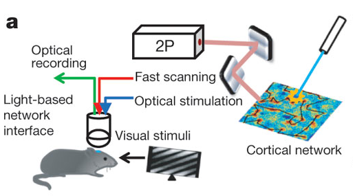
We published another double header yesterday, this time on the role of particular cell types in visual responses. Both studies describe the effect of optogenetically manipulating various interneuron classes in mouse visual cortex. The papers are Lee et al. from Yang Dan‘s lab and Wilson et al. from Mriganka Sur‘s labs. And in fact, both were preceded by Atallah et al. from Massimo Scanziani’s lab, which appeared in Neuron earlier this year. Which means a bonanza of data on the effects of activating parvalbumin-expressing interneurons, and also a bonanza of different conclusions about their exact role – everyone comes to slightly different conclusions.

We’ve discussed joint (and triple) publication a number of times already on this blog, including situations where findings diverge. We even just recently discussed a triple publication involving a paper from Yang Dan’s lab. So I’ll leave it to you to extrapolate the editorial discussions that likely took place in this case, but if anyone wants to know more, leave a comment. Instead, I’ll touch on another question that we get asked fairly regularly: what do we do when authors submit papers to us in quick succession? Is there a limit on how many papers from one lab we will publish per year? Since we’re mentioning today a paper by an author who had a paper covered in the previous blog post, you can infer that number is at least two. Just kidding. Of course we have no limit. Scientific progress unfolds at different rates, and sometimes labs have some very good years. As long as a study has potential impact, we are happy to consider. Continue reading

A tale of three papers

From Figure 1, Li et al.

I wanted the title of this post to be “A tale of two one two three papers” but I couldn’t figure out how to get strikethroughs in the title field. And I thought “A tale of two, make that one, no make that two again, oops now three” might be a bit cumbersome. As promised, here’s another installment of the discussion of what happens when we receive conceptually related/overlapping papers. It starts with a paper that appeared just yesterday in Neuron by Kenichi Ohki and colleagues describing how mouse visual cortex neurons that developed from the same neural progenitor cell tend to be more similar functionally than those that did not.

Why is this significant? First a little background. Cells in visual cortex are tuned to different aspects of visual stimuli, such as orientation or direction, and anatomically are organized quite specifically. Cells with similar preferences tend to cluster together and to be selectively connected with each other (though to differing degrees in different species), and this specificity may underlie some of the many computations required to turn photons of light hitting our eyes into comprehensible percepts.  It’s been proposed that this clustering could start in early development; neurons born from the same neural progenitor migrate vertically to form columns of sibling neurons, and could be the basis for clusters of adult cells with similar properties. That link hasn’t been demonstrated experimentally until now, and Ohtsuki et al. provides some evidence in support of it.

Now, visual cortex aficionados among you may think this sounds a bit similar to Li et al., a paper by Yang Dan and colleagues that appeared a few months ago, and indeed it is. And you may also recall that THAT paper appeared alongside Yu et al. from Songhai Shi’s lab about the development of synapses between sibling neurons.

So here’s the story from the beginning (or rather, the beginning of our involvement with the manuscripts).

Continue reading

Autism, synapses and mice – pairs division

From Won et al.

Again, we’re behind on blogging – you guys are keeping us busy with great neuroscience – but here is the story of a pair of papers that appeared back to back in last week’s issue and a continuation of the discussion started here by Noah about the process of joint publication. The two papers by Tobias Boeckers and colleagues and by Eunjoon Kim and colleagues were independently submitted and both describe autism-like phenotypes of mice with mutations in the gene Shank2. In human studies, SHANK2 has been associated with rare cases of autism and these two mice add to the ever-growing list of rodents (according to SFARI.org, 17 rodent models debuted in 2011 alone) that are being created to investigate the functional consequences of genetic mutations linked to autism, in the hopes of understanding mechanisms underlying core symptoms. Shank2 is a scaffolding protein that regulates excitatory synapse function by holding together various molecules such as neurotransmitter receptors and signaling proteins. Mutations in another member of the same gene family, SHANK3, are also associated with human autism, and mutant mice display behaviors reminiscent of ASD symptoms, such as social deficits and obsessive behavior. So this protein family, and more generally, glutamatergic transmission, is potentially one promising line of investigation. Continue reading

Positive feedback drives network (and manuscript) maturation.

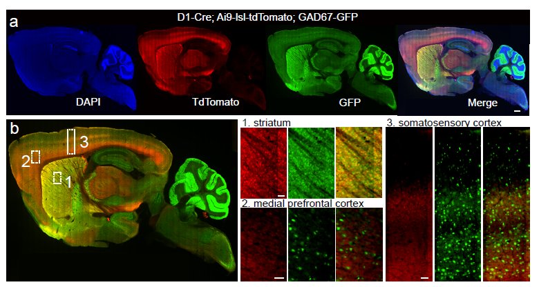
Whole-brain anatomical mapping of D1-Cre expression in inhibitory neurons (from Supp Fig.2)

It really is an embarrassment of riches here at Nature these days, what with so many excellent neuroscience-related studies emerging. Just in the last couple of weeks, we’ve had the following studies:

So really, a lot to write about from a science perspective. However, this blog is dedicated to bringing you the editorial back-story, so I wanted to touch on yet another interesting study, published in print today. This new paper offers an opportunity to discuss an important editorial issue: the manuscript appeal process. For more details, you can always read the appropriate section in our guide to authors. But it’s often helpful to follow a particular [successful] example in order to illustrate the process. Continue reading

“There is no spoon…”: Paralyzed fish navigates virtual environment while we watch its brain

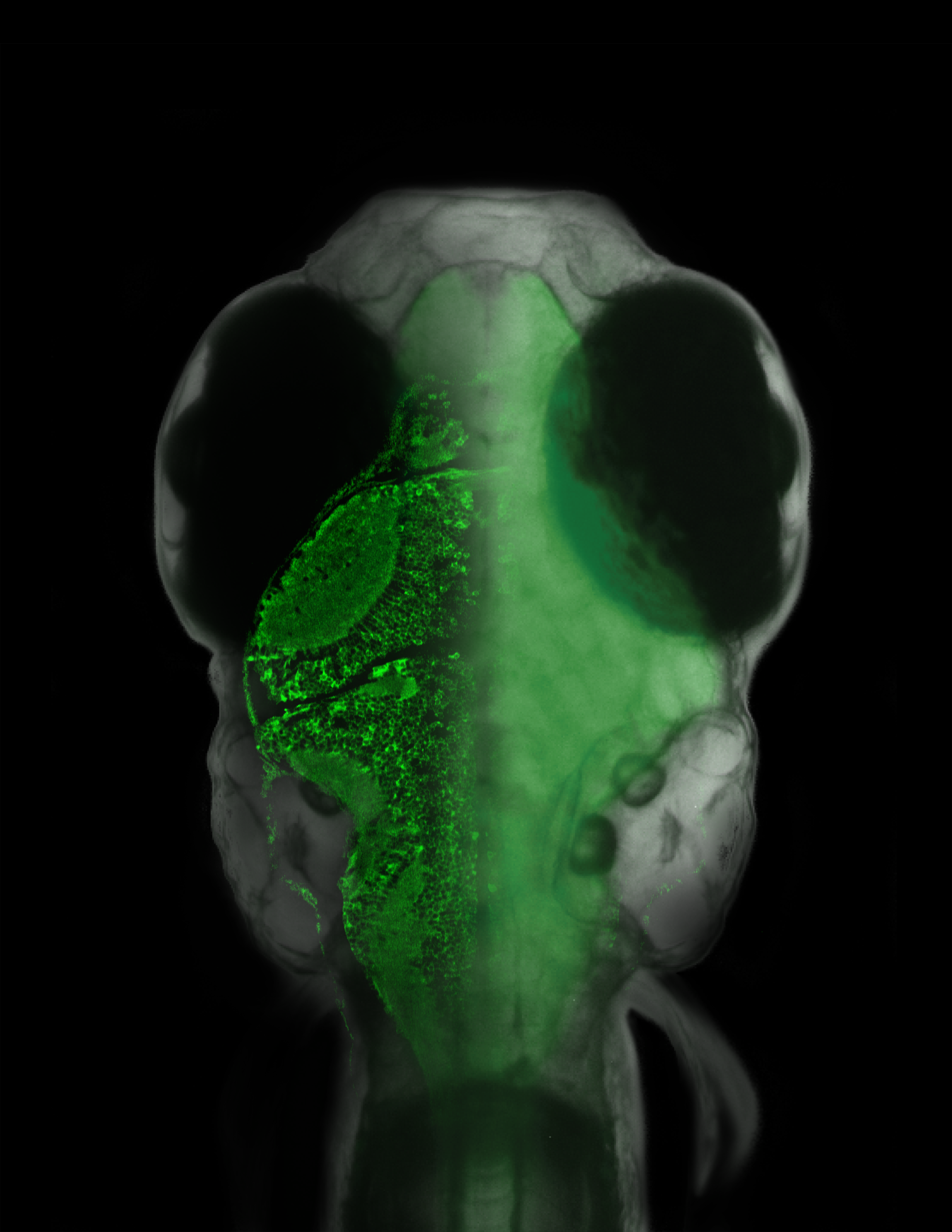
Overlaid on the micrograph of the fish is a slice of its brain measured with a laser scanning microscope, in which single neurons are visible.{credit}(courtesy of Ahrens et al.){/credit}

Sometimes an experiment will just reach off the page and slap you in the face, demanding attention. This happens to me every so often and I must admit, our latest paper from the lab of Florien Engert induced such an experience. There have been several cool, technical tours-de-force (is that proper grammar??) over the last few years involving different creatures navigating in a virtual environment while neuronal activity was monitored. These include a mouse running on a spherical treadmill, as well as a fly marching along a similar treadmill-style ball. But in these examples, having the subject head-fixed (for the stability of recordings in the brain, either with electrodes or through imaging) was moderately non-intrusive since walking motions were independent of the head. The same can’t be said for the subject in this latest example of a virtual reality navigator: a wriggling, swimming fish. Therefore, a more creative solution had to be sought and in a paper published online yesterday, Ahrens, Engert and colleagues decided that paralysis was the way to go in order to follow the neural activity of this navigating fish. Continue reading

Open access and criminal minds: Boston events this week

A pick of events for this week. See calendar for details.

Monday

“Does mandating free online access to papers resulting from federally funded research violate the Copyright Act or treaty obligations? “ Find out Monday when Mark Seeley, VP and General Counsel of Elsevier, and Peter Suber, of the Berkman Center for Internet and Society meet to discuss the issue. They promise to cover open access mandates including the pending Federal Research Public Access Act, as well as the National Institutes of Health Public Access Policy. Click here for the live webcast from Harvard.

Tuesday

Frances Chew, Professor of Ecology at Tufts University The Cambridge Entomological Club speaks on the return of the mustard white butterfly, which had been endangered by “serial invasions of garlic mustard, parasitoid biological control agents for the related cabbage white butterfly, and other exotic plant species.”  The threatened butterfly is now poised to recover, “an unintended consequence of recent species introductions.”

Wednesday

Seeing the forest AND the trees: Modeling ecosystem climate.  Harvard post doc Naomi Marcil Levine will describe an model that identifies “ecological mechanisms that provide tropical forests with resiliency to changes in climate.”

Thursday

Four experts — including  defense lawyer F. Lee Baily and former Harvard provost Stephen Hyman —  discuss “Neuroscience and the Criminal Mind” at the Starr Center in Boston.