Top models

Speaking of mammals (see the end of my previous entry), not even rats and mice always cut it when it comes to providing good models of human disease. Take, for example, cystic fibrosis. There are a couple of mouse models of the disease (we have published at least one of them), but the community does not seem to be satisfied with them. It is therefore great to see a pair of papers in the JCI reporting on two new attempts at generating the ideal model of cystic fibrosis.

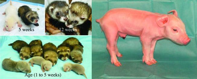
The two of studies are very similar. In the first one, Xingshen Sun and colleagues report the first description of genetically engineered ferrets. They started by targetting the CFTR gene (the gene affected in the disease) in fibroblasts using an adeno-associated viral (AAV) construct, and then used a nuclear transfer protocol to obtain cloned ferrets heterozygous for the CFTR mutation. In the second one, Christopher Rogers and colleagues employed a similar strategy in pigs to obtain heterozygous piglets carrying the CFTR mutation.

The next steps will be to establish how much these models truly recapitulate human disease, and then use them to learn new biology about the disease and/or for preclinical drug-discovery work.

The figures, taken from the papers, show the cloned CFTR ferrets and a non-cloned albino at different ages, and the first CFTR heterozygous pig at one day of age.

ferrets.JPG

Retinoids, skincare and Matthew Wood

People with acne will find this JCI paper of interest. 13-cis retinoic acid can be used to treat acne, as it can kill human sebaceous-gland cells by apoptosis. The molecule is teratogenic, though, making it necessary to look for alternatives. As the mechanism of action of 13-cis retinoic acid is unknown, Amanda Nelson and her colleagues tried to elucidate it, hoping to identify new targets for the treatment of the bothersome skin condition. Using transcriptional profiling of skin cells from people with acne and cultured sebaceous glands, they found that lipocalin-2 was distinctively upregulated by treatment with 13-cis retinoic acid. They also found that the apoptotic effect of 13-cis retinoic acid indeed depended on the expression of neutrophil gelatinase–associated lipocalin (NGAL), the protein encoded by lipocalin-2; by using siRNA to lipocalin-2, they blocked the apoptotic effect, and by adding recombinant NGAL, they promoted it. It is therefore conceivable that manipulating NGAL expression could lead to a new way to fight acne.

acne.jpg

A more serious pathology with a connection to retinoids is Matthew-Wood syndrome, a fatal disease characterized by multisystem developmental malformations that has been linked to mutations in STRA6. STRA6 interacts with retinol-binding protein 4 (RBP4), which is, in turn, a carrier of retinoids (vitamin A and its derivatives). A paper published in Cell Metabolism establishes that the biochemical interaction between STRA6 and RBP4 is indeed functionally relevant. Studying zebrafish embryos, Andrea Isken and colleagues found that Stra6 deficiency allows more Rbp4 to remain free and to carry an excess amount of retinoids to several embryonic tissues, including bone, heart and eye. In fact, reducing the levels of Rbp4 prevented these effects. The findings provide a nice molecular account of Matthew-Wood syndrome, although I cannot help but wish that the authors had done the in vivo experiments in a mammal. I must confess that, when we evaluate papers at Nature Medicine, we’re seldom enthused by data from zebrafish, Drosophila or C. elegans, as the relevance of these models to human physiology tends to be harder to ascertain. Nothing personal against the fish or the invertebrates, though.

zebrafish.jpg

Understanding aging

Three papers published this past Sunday touch upon different aspects of the aging problem. The first one appeared in Nature and is authored by Rui Yi and colleagues, who found that microRNA-203 promotes the differentiation of skin stem cells by repressing “stemness”. In stratified epithelia, stem cells located basally are crucial for self renewal. As these cells leave the basal zone, they differentiate and cease to behave like stem cells. What the authors found is that microRNA-203 is crucial for this differentiation process, leading the stem cells to exit the cell cycle. Mechanistically, this effect depends on repression of p63 expression, a molecule that had previously been shown to regulate stem-cell maintenance in epithelia.

wrinkles.jpg

The second one is on one of my favorite topics — progeria. Writing in Nature Cell Biology, Paola Scaffidi and Tom Misteli report that expression of mutant lamin-A, the molecule that causes Hutchinson-Gilford Progeria Syndrome (HGPS), interferes with the function of human mesenchymal stem cells (hMSCs) by promoting the activation of downstream

effectors of Notch, affecting the differentiation potential of hMSCs. The in vivo relevance of these results to HGPS and to normal aging remains to be established, but the possibility is indeed tantalizing.

skin.jpg

The third one is a Brief Communication in Nature Genetics. In it, Marc Vermulst and his colleagues establish a link between mitochondrial DNA (mtDNA) deletions and aging in the so-called Polga mice, which harbor a proofreading-deficient copy of polymerase gamma and are characterized by premature aging. They found that the rate at which different tissues accumulate mtDNA mutations before they reach phenotypic expression differs profoundly — brain, heart and gut are among the most affected parts of the body. The question remains, though, if these mtDNA mutations are also relevant during normal aging in wild-type mice and, of course, in humans.

Two on ALS

Even though my background is in neuroscience, I rarely write about this topic. But wo papers on amyotrophic lateral sclerosis (ALS) from the latest issue of the Journal of Neuroscience struck me as interesting to talk about.

In the first one, Fiona Laird and her colleagues generated transgenic mice that express wild-type and mutant forms of the human protein dynactin p150-Glued. As mutant forms of this molecule had been linked to ALS, they decided to explore the mechanism whereby dynactin p150-Glued contributes to the pathology. They found that expression of dynactin p150-Glued carrying a mutation that had been linked to the disease in patients led to motor neuron disease in transgenic mice, something that was not seen in mice overexpressing the wild-type form of the human protein.

The paper is very nice in that it provides a very detailed account of the neuropathology the authors see in the mouse, including some intriguing evidence of autophagic cell death. The picture below, which comes from the paper, is a silver-stained section of the spinal cord from a mutant mouse, showing dark, presumably dying, motor neurons (arrowheads) that are not seen in control mice. Unfortunately, the authors didn’t get to explore the hardcore molecular mechanisms that account for the motor neuron death. But they now have a useful system to ask more mechanistic questions to understand the role of dynactin p150-Glued in cell death and investigate its actual relationship to human ALS.

laird.JPG

The second study deals with a question that has occupied the field for some time. We know that mutations in superoxide dismutase (SOD) are linked to familial forms of ALS, but where does SOD need to be expressed to cause disease: in neurons, in glia, in muscle? Dick Jaarsma and his colleagues tried to get at this question by generating transgenic mice that expressed mutant SOD only in neurons. The figure below, from the original paper, shows spinal cord sections from mice that expressed the mutant protein only in neurons (top left and bottom right) or ubiquitously (top middle).

jaarsma.JPG

This is not the first time that neuron-specific expression of SOD has been tried, but it is perhaps the first time in which it is found to effectively kill the motor neurons. In other words, these findings fly in the face of other studies reporting no motor neuron death in mice with neuron-specific expression of mutant SOD and of papers specifically identifying a contribution of extraneuronal SOD to ALS. Not unexpectedly, there is at present no definitive way to reconcile these disparate observations, other than invoking technical differences in the studies or stating that the cell-autonomous effect reported by Jaarsma et al. does not negate an additional contribution from glial SOD. What we can say for sure is that we don’t yet understand the neuron/glia/muscle interplay in ALS, and that it will be quite hard to establish if the contributions of mutant SOD from each of these sources in transgenic mice are indeed relevant to the human condition.

Times of change for prostate cancer

As I approach the age at which the word ‘prostate’ starts sounding like a funereal drum, I become more interested in studies such as those published this week in the NEJM and about three weeks ago in Nature Genetics.

The NEJM paper, by Lilly Zheng and colleagues, shows that single-nucleotide polymorphisms (SNPs) in five chromosomal regions, each of which had previously and independently been associated with prostate cancer, have a cumulative association with the disease when considered in combination. The authors estimate that the five SNPs and a family history of prostate cancer account for as many as 46% of the cases in the Swedish population they studied.

The three Nature Genetics papers, by Julius Gudmundsson et al., Gilles Thomas et al. and Rosalind Eeles et al., all of which are nicely summarized in the journal’s March editorial, disclose multiple new susceptibility loci associated with prostate cancer that, together with other loci identified in 2006 and 2007, give us plenty of new avenues to explore in order to understand the disease.

The most immediate implication of findings of this sort is often diagnostic — if you identify gene variants that are linked to a disease, you can ask questions about how good these variants are at predicting onset and/or progression of the pathology. Validating the diagnostic value of these genomic data often requires blinded samples analyzed in a prospective (preferably longitudinal) fashion.

The findings could also help us understand the biology of the disease, although this almost always takes more time and is not always pursued, as it is very challenging: you need to identify with precision the protein whose gene harbors the relevant SNP, then establish how the SNP affects protein function, and finally look at how this altered function modifies the physiology of the cell as it becomes tumorigenic in an in vivo setting.

This is what we at Nature Medicine look for when we evaluate submissions that report new associations of SNPs or mutations with disease, which is why we don’t tend to publish too many of these kind of studies. That said, these ruminations do not take anything from the value of these four studies, which shine some more light on the black box that prostate cancer has turned out to be.

Michael’s insight and beyond

In addition to the Insight on Cardiovascular Disease, edited by Nature Medicine‘s own Michael Basson, a couple of papers caught my attention from this past Thursday’s issue of Nature.

First, the analysis of multiple sclerosis (MS) lesions by laser-capture microdissection and proteomics, which led May Han and colleagues to identify two potential therapeutic targets for the disease — tissue factor and protein C inhibitor — both of which participate during coagulation. Indeed, the authors went on to show that blocking the action of thrombin (which signals downstream of tissue factor) or administering activated protein C (to counter the increased levels of its inhibitor) ameliorated pathology in an animal model of MS. The image below, from the Nature paper, shows astrogliosis in a chronic MS plaque, revealed by and anti-GFAP antibody.

steinman.JPG

Second, the discovery by Xiaoyong Yang and colleagues of a link between O-GlcNac transferase and insulin resistance. We already knew that glucose flux through the hexosamine biosynthetic pathway leads O-GlcNac transferase to attach the sugar O-linked beta-N-acetylglucosamine (O-GlcNac) to proteins, thereby acting as a nutrient sensor. The new study shows that O-GlcNac transferase has a binding site for phosphatidylinositol 3,4,5-trisphosphate (PIP3), a key mediator of insulin signaling. Upon binding, PIP3 recruits O-GlcNac transferase to the plasma membrane, where it sticks O-GlcNac to proteins of the insulin signaling pathway, reducing their responsiveness to insulin (see the figure below, which I borrowed from the paper; O-GlcNac transferase is labeled as OGT). In vivo, liver overexpression of O-GlcNac transferase causes insulin resistance, pointing to the likely functional relevance of this mechanism.

evans.JPG

Mutants, revertants and innate responders

Two sets of papers caught my attention over the past couple of days. Apologies if they are old hat for those of you who work in these fields. It’s hard to keep up with all the ToC alerts I get.

The first is a doublet from Nature on the mechanism whereby certain tumors acquire resistence to chemotherapy. The studies, by Wataru Sakai and colleagues and by Stacey Edwards and colleagues focused on tumors that carry mutations in BRCA2 and are therefore sensitive to platinum compounds like cisplatin. In some cases, these tumors develop resistance to cisplatin, and what both studies show is that the development of resistance depends on the appearance of new mutations in BRCA2, which restore the open reading frame of the protein. Although this is perhaps not incredibly surprising, particularly because similar secondary mutations had been observed in cases of resistance to imatinib in leukemia, this finding has obvious clinical implications for people who become unresponsive to cisplatin.

The other paper, by Marielle Gold and her colleagues in PLoS Pathogens, reports on the existence of a novel population of human T cells that innately recognize Mycobacterium tuberculosis (Mtb). These cells, which the authors isolated from newborns who were very unlikely to have ever been exposed to the bacterium, exist at relatively high frequencies and respond to Mtb-infected cells by producing IFN-γ. The authors assert that this is the first demonstration of a human innate pathogen-specific T cell and refer to preliminary experiments showing that other thymocytes can also respond to other pathogens including Staphylococcus aureus and Escherichia coli. How this innate recognition comes about in the fisst place strikes me as a pretty interesting question for follow-up studies.

Something for the weekend

A couple of days ago I was saying that the problem with blogging (at least for me) is lack of discipline. So I figured that one way to become a bit more disciplined, and hopefully post stuff that people will find of interest, would be to write a brief entry every time I come across a paper that I think is particularly interesting. I’m calling this category of entries “Journal club” for lack of a better name, as I don’t think I want to (nor could) write an extensive critique of the paper in question. Instead, the purpose of doing this is to flag a paper as something that is of interest to an editor of Nature Medicine, and let those of you who work in the relevant field do the detailed evaluation of the contribution.

To get things rolling, here’s three papers:

1) A study by Stephen Hauser and his colleagues in the NEJM reports that rituximab, a drug used for the treatment of non-Hodgkin lymphoma and rheumatoid arthritis, could also be useful to treat multiple sclerosis. Their clinical trial involved 104 people, 69 of whom received two one-gram doses of the drug (which acts by depleting CD20+ B cells). The trial lasted 48 weeks and showed a reduction in the number of inflammatory brain lesions and clinical relapses in the treated patients versus the controls over this time period. Although the trial wasn’t designed to establish long-term safety or efficacy, it is indeed promising for people with MS.

2) In Immunity, Jackson Egen and his colleagues report on their use of high-resolution multiplex static imaging and intravital multiphoton microscopy to give us an unprecedented look at granulomas — masses of inflammatory cells that arise owing to the persistence of an infectious agent in host tissue and that are critical for host protection.

Granulomas, which are often seen in people with tuberculosis, contain different cell types including lymphocytes, macrophages and fibroblasts. In their study, the authors found that, after infection with Mycobacterium bovis, Kupffer cells in the liver capture circulating bacteria and subsequently form the nucleus of a new granuloma by recruiting uninfected liver-resident macrophages and blood-derived monocytes. Within the granuloma, these cells set up an immobile matrix that attracts a dynamic population of T cells in a TNF-alpha-dependent manner. You ought to check out their movies.

3) To continue with the microbiology theme and the topic of the interaction of bacteria with host tissue, Science just published a study on the mechanism whereby tissue abscesses can inhibit bacterial growth. Brian Corbin and his colleagues found that calprotectin — a neutrophil-derived protein — can stall the growth of Staphylococcus aureus inside an abscess. Mechanistically, the effect of calprotectin depended on its ability to chelate Zn2+ and Mn2+, thereby interfering with the transcriptional machinery of the bacterium. In vivo, mice lacking calprotectin had abscesses with higher levels of metals that seemed to favor staphylococcal proliferation. Whether metal chelation can work as a general strategy to inhibit bacterial growth inside an abscess remains to be seen, but the possibility is certainly tantalizing.