Optogenetics in neuroscience at Nature Methods

The optogenetic manipulation of cellular properties has not only revolutionized neuroscience, but this technology can also be applied to the manipulation of signaling pathways, transcription or other processes in non-neuronal cells. Here, we highlight some of the papers we have published on the neuroscience side of optogenetics.

Optogenetic tools

2014 has been an exciting year for us with the publication of new optogenetic tools. Klapoetke and Boyden developed Chrimson and Chronos, two channelrhodopsins that they discovered in a screen of algal transcriptomes. Chrimson is more red-shifted than previously known channelrhodopsins while Chronos has faster kinetics. Hochbaum and Cohen described another algal channelrhodopsin called CheRiff, which is highly sensitive to blue light stimulation, making it compatible with red-shifted voltage sensors.

Previously, we published papers describing modifications to optogenetic tools. For example, Prakash and Deisseroth tailored opsin with custom properties. To ensure stoichiometric expression of optogenetic activators and/or inhibitors, Kleinlogel and Bamberg simply and elegantly fused the two proteins into a single chain. Depending on the two partners, this marriage can lead to synergisms or bidirectional effects. Finally, Mattis and Deisseroth undertook a comprehensive characterization of available tools.

Optogenetic applications

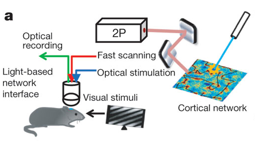
Since the initial description of Channelrhodopsin2 (ChR2) as an efficient tool to evoke neural activity in a light-dependent manner, we have seen a flurry of papers applying ChR2 for a variety of questions in neuroscience. For instance, Zhang and Oertner combined this tool with two-photon calcium imaging in rat slices to study synaptic plasticity. Liewald and Gottschalk applied the same methodology to analyze synaptic function in freely moving C. elegans.

ChR2 can also be used to map the function of brains regions as Ayling and Murphy demonstrated by evoking activity in limb muscles via light stimulation in the motor cortex of ChR2 transgenic mice. Similarly, Guo and Ramanathan mapped neural circuitry in C. elegans by combining ChR2-mediated neural activation with imaging of a genetically encoded calcium sensor in downstream neurons. To facilitate circuit mapping in mice, Zhao and Feng generated mouse lines that express ChR2 in GABAergic, cholinergic, serotonergic or parvalbumin-expressing neurons.

While ChR2 is a very popular tool in optogenetics, other family members can do the job as well. C1V1T is a fusion of two different opsins and is particularly useful when applying two-photon excitation, as shown by Packer and Yuste. ReaChR is activated by red light and thus especially useful in vivo. Inagaki and Anderson studied courtship behavior in Drosophila with this tool.

Method of the Year

We celebrated the impact of optogenetics by recognizing the technology as our Method of the Year 2010. We marked the occasion with the publication of special Commentaries on the subjects. Deisseroth discussed the past, present and future of optogenetics. Hegemann and Möglich deliberate on the exploration of new optogenetic tools. And Peron and Svoboda illuminated us on the precise delivery of optogenetic stimulation. In addition, our News Feature recounted the stories behind the “Light tools”.

If we have sparked your interest, the mentioned papers are listed below.

We are excited to hear about the upcoming developments in optogenetics from you.


Nathan C Klapoetke, Yasunobu Murata, Sung Soo Kim, Stefan R Pulver, Amanda Birdsey-Benson, Yong Ku Cho, Tania K Morimoto, Amy S Chuong, Eric J Carpenter, Zhijian Tian, Jun Wang, Yinlong Xie, Zhixiang Yan, Yong Zhang, Brian Y Chow, Barbara Surek, Michael Melkonian, Vivek Jayaraman, Martha Constantine-Paton, Gane Ka-Shu Wong & Edward S Boyden
Independent optical excitation of distinct neural populations
Nature Methods 11, 338–346 (2014) doi:10.1038/nmeth.2836

Daniel R Hochbaum, Yongxin Zhao, Samouil L Farhi, Nathan Klapoetke, Christopher A Werley, Vikrant Kapoor, Peng Zou, Joel M Kralj, Dougal Maclaurin, Niklas Smedemark-Margulies, Jessica L Saulnier, Gabriella L Boulting, Christoph Straub, Yong Ku Cho, Michael Melkonian, Gane Ka-Shu Wong, D Jed Harrison, Venkatesh N Murthy, Bernardo L Sabatini, Edward S Boyden, Robert E Campbell & Adam E Cohen
All-optical electrophysiology in mammalian neurons using engineered microbial rhodopsins
Nature Methods 11, 825–833 (2014) doi:10.1038/nmeth.3000

Rohit Prakash, Ofer Yizhar, Benjamin Grewe, Charu Ramakrishnan, Nancy Wang, Inbal Goshen, Adam M Packer, Darcy S Peterka, Rafael Yuste, Mark J Schnitzer & Karl Deisseroth
Two-photon optogenetic toolbox for fast inhibition, excitation and bistable modulation
Nature Methods 9, 1171–1179 (2012) doi:10.1038/nmeth.2215

Sonja Kleinlogel, Ulrich Terpitz, Barbara Legrum, Deniz Gökbuget, Edward S Boyden, Christian Bamann, Phillip G Wood & Ernst Bamberg
A gene-fusion strategy for stoichiometric and co-localized expression of light-gated membrane proteins
Nature Methods 8, 1083–1088 (2011) doi:10.1038/nmeth.1766

Joanna Mattis, Kay M Tye, Emily A Ferenczi, Charu Ramakrishnan, Daniel J O’Shea, Rohit Prakash, Lisa A Gunaydin, Minsuk Hyun, Lief E Fenno, Viviana Gradinaru, Ofer Yizhar & Karl Deisseroth
Principles for applying optogenetic tools derived from direct comparative analysis of microbial opsins
Nature Methods 9, 159–172 (2012) doi:10.1038/nmeth.1808

Yan-Ping Zhang & Thomas G Oertner
Optical induction of synaptic plasticity using a light-sensitive channel
Nature Methods 4, 139 – 141 (2006) doi:10.1038/nmeth988

Jana F Liewald, Martin Brauner, Greg J Stephens, Magali Bouhours, Christian Schultheis, Mei Zhen & Alexander Gottschalk
Optogenetic analysis of synaptic function
Nature Methods 5, 895 – 902 (2008) doi:10.1038/nmeth.1252

Oliver G S Ayling, Thomas C Harrison, Jamie D Boyd, Alexander Goroshkov & Timothy H Murphy
Automated light-based mapping of motor cortex by photoactivation of channelrhodopsin-2 transgenic mice
Nature Methods 6, 219 – 224 (2009) doi:10.1038/nmeth.1303

Zengcai V Guo, Anne C Hart & Sharad Ramanathan
Optical interrogation of neural circuits in Caenorhabditis elegans
Nature Methods 6, 891 – 896 (2009) doi:10.1038/nmeth.1397

Shengli Zhao, Jonathan T Ting, Hisham E Atallah, Li Qiu, Jie Tan, Bernd Gloss, George J Augustine, Karl Deisseroth, Minmin Luo, Ann M Graybiel & Guoping Feng
Cell type–specific channelrhodopsin-2 transgenic mice for optogenetic dissection of neural circuitry function
Nature Methods 8, 745-752 (2011) doi:10.1038/nmeth.1668

Adam M Packer, Darcy S Peterka, Jan J Hirtz, Rohit Prakash, Karl Deisseroth & Rafael Yuste
Two-photon optogenetics of dendritic spines and neural circuits
Nature Methods 9, 1202–1205 (2012) doi:10.1038/nmeth.2249

Hidehiko K Inagaki, Yonil Jung, Eric D Hoopfer, Allan M Wong, Neeli Mishra, John Y Lin, Roger Y Tsien & David J Anderson
Optogenetic control of Drosophila using a red-shifted channelrhodopsin reveals experience-dependent influences on courtship
Nature Methods 11, 325–332 (2014) doi:10.1038/nmeth.2765

Karl Deisseroth
Optogenetics
Nature Methods 8, 26–29 (2011) doi:10.1038/nmeth.f.324

Peter Hegemann & Andreas Möglich
Channelrhodopsin engineering and exploration of new optogenetic tools
Nature Methods 8, 39–42 (2011) doi:10.1038/nmeth.f.327

Simon Peron & Karel Svoboda
From cudgel to scalpel: toward precise neural control with optogenetics
Nature Methods 8, 30–34 (2011) doi:10.1038/nmeth.f.325

Monya Baker
Light tools
Nature Methods 8, 19–22 (2011) doi:10.1038/nmeth.f.322

Is phototoxicity compromising experimental results?

Light-induced damage to biological samples during fluorescence imaging is known to occur but receives too little attention by researchers.

The December Technology Feature in Nature Methods asks if super-resolution microscopy is right for you and a point that comes up repeatedly from the researchers we interviewed is the danger of phototoxicity and photodamage caused by the high irradiation intensities needed for the illuminating light. This has long been a concern with these methods and many of the papers describing them mention it.

But as discussed in the December Editorial, even fluorescence microscopy with low irradiation intensities can cause dangerous levels of phototoxicity that permanently damage the sample. Microscopists are aware of these concerns but there has been little effort to implement processes intended to reduce the likelihood of it compromising research study results. Dave Piston, Director of the Biophotonics Institute at Vanderbilt University School of Medicine, laments that while phototoxicity is a big deal he has gotten zero traction with NIH reviewers on trying to build some rules for it.

There are some good resources available to researchers that highlight the dangers of phototoxicity and provide advice on how to limit it. Methods in Cell Biology Vol 114 has an excellent chapter by Magidson and Khodjakov, Circumventing Photodamage in Live-Cell Microscopy, that should be mandatory reading for all researchers using fluorescence microscopy for biological research. Also, Nikon’s MicroscopyU has a literature list with several dozen references and recommended reading on phototoxicity. It could use some updating but is still useful.

Despite the amount of microscopy literature that discusses phototoxicity, discussion of the phenomenon in research articles published in Nature Journals is conspicuously absent. This is highlighted by a simple full-text search we performed on the HTML versions of research articles published in Nature, Nature Cell Biology, Nature Immunology, Nature Methods and Nature Neuroscience. The articles retrieved were limited to original research articles.

The table below lists the number of occurrences of each of the listed words in the period from January 1, 2005 to November 3, 2013 in each of the indicated journals. The percentages represent the number fraction of articles containing ‘phototoxicity’ relative to the numbers of articles containing each of the microscopy- or fluorescence-related terms. Note that this is NOT a measure of co-occurrence, only a measure of how common the term ‘phototoxicity’ is relative to the other terms.

phototoxicity fluorescence fluorescent microscopy microscope
# # % # % # % # %
Nature 8 2120 0.4% 1925 0.4% 1995 0.4% 1918 0.4%
Nature Cell Biology 8 815 1.0% 728 1.1% 866 0.9% 822 1.0%
Nature Immunology 6 552 1.1% 574 1.0% 408 1.5% 326 1.8%
Nature Methods 27 565 4.8% 494 5.5% 441 6.1% 407 6.6%
Nature Neuroscience 18 639 2.8% 727 2.5% 587 3.1% 736 2.4%


The same analysis was repeated with the term ‘photodamage’ to determine if there was a substantial difference in the usage of these two similar terms.

photodamage fluorescence fluorescent microscopy microscope
# # % # % # % # %
Nature 18 2120 0.8% 1925 0.9% 1995 0.9% 1918 0.9%
Nature Cell Biology 6 815 0.7% 728 0.8% 866 0.7% 822 0.7%
Nature Immunology 2 552 0.4% 574 0.3% 408 0.5% 326 0.6%
Nature Methods 29 565 5.1% 494 5.9% 441 6.6% 407 7.1%
Nature Neuroscience 12 639 1.9% 727 1.7% 587 2.0% 736 1.6%


These results carry the potentially large caveat that the analysis did not include the text of the supplementary information, but the rarity with which phototoxicity or photodamage is discussed (0.4% to 7% relative to microscopy terms) suggests that researchers don’t appreciate how important it is to pay attention to artifacts that result from light irradiation. Luckily, there are exceptions to this state of affairs.

An excellent example of testing for phototoxicity and the subtle effects it can induce can be found in a manuscript from Jeff Magee’s lab at Janelia Farm Research Campus published last year in Nature. Quoting from the manuscript, “Particular care was taken to limit photodamage during imaging and uncaging. This included the use of a passive 8× pulse splitter in the uncaging path in most experiments to reduce photodamage drastically [Ji, N. et al. Nat. Methods (2008)]. Basal fluorescence of both channels was continuously monitored as an immediate indicator of damage to cellular structures. Subtle signs of damage included decreases in or loss of phasic Ca2+ signals in spine heads in response to either uncaging or current injection, small but persistent depolarization following uncaging, and changes in the kinetics of voltage responses to uncaging or current injection. Experiments were terminated if neurons exhibited any of these phenomena.”

It is easy to see how these changes in Ca2+ responses could easily have been interpreted as real biological effects caused by the uncaged glutamate, rather than the uncaging light itself.

It is unrealistic to expect that any mandates or oversight would be able to prevent or detect such consequences of phototoxicity in research studies. It is essential that investigators themselves be vigilant and implement appropriate controls to detect these effects. Na Ji, also at Janelia Farm Research Campus says, “It is not enough to only look for instant and dramatic signs of phototoxicity. Sometimes the effects may be more subtle and even unperceivable during the imaging period, but may become obvious when the same sample is imaged the next day. Care has to be taken in data collection and interpretation, especially when the biological process under investigation itself is a subtle one.”

Finally, the application is just as important as the imaging method being used. For example, light-sheet microscopy is excellent at reducing irradiation levels in volumetric imaging. But some applications of super-resolution microscopy, even on living samples, might be less susceptible to artifacts caused by phototoxicity than are sensitive long-term imaging applications of living samples by light-sheet microscopy. Nobody’s microscope earns them a free pass on the dangers of photodamage arising from phototoxicity. Everyone needs to be vigilant.

Update: A reader helpfully pointed out that the danger of phototoxicity and photodamage also applies to optogenetics, where light (often in the blue region of the spectrum) is used to control protein activity.

Too much of a good thing?

From Wilson et al.

We published another double header yesterday, this time on the role of particular cell types in visual responses. Both studies describe the effect of optogenetically manipulating various interneuron classes in mouse visual cortex. The papers are Lee et al. from Yang Dan‘s lab and Wilson et al. from Mriganka Sur‘s labs. And in fact, both were preceded by Atallah et al. from Massimo Scanziani’s lab, which appeared in Neuron earlier this year. Which means a bonanza of data on the effects of activating parvalbumin-expressing interneurons, and also a bonanza of different conclusions about their exact role – everyone comes to slightly different conclusions.

We’ve discussed joint (and triple) publication a number of times already on this blog, including situations where findings diverge. We even just recently discussed a triple publication involving a paper from Yang Dan’s lab. So I’ll leave it to you to extrapolate the editorial discussions that likely took place in this case, but if anyone wants to know more, leave a comment. Instead, I’ll touch on another question that we get asked fairly regularly: what do we do when authors submit papers to us in quick succession? Is there a limit on how many papers from one lab we will publish per year? Since we’re mentioning today a paper by an author who had a paper covered in the previous blog post, you can infer that number is at least two. Just kidding. Of course we have no limit. Scientific progress unfolds at different rates, and sometimes labs have some very good years. As long as a study has potential impact, we are happy to consider. Continue reading

Positive feedback drives network (and manuscript) maturation.

Whole-brain anatomical mapping of D1-Cre expression in inhibitory neurons (from Supp Fig.2)

It really is an embarrassment of riches here at Nature these days, what with so many excellent neuroscience-related studies emerging. Just in the last couple of weeks, we’ve had the following studies:

So really, a lot to write about from a science perspective. However, this blog is dedicated to bringing you the editorial back-story, so I wanted to touch on yet another interesting study, published in print today. This new paper offers an opportunity to discuss an important editorial issue: the manuscript appeal process. For more details, you can always read the appropriate section in our guide to authors. But it’s often helpful to follow a particular [successful] example in order to illustrate the process. Continue reading

Fear of the Light

fear-of-the-light**PLEASE SEE UPDATES BELOW**

It is commonly believed that distinct mini-networks of neurons, firing together, may be the means by which memories and other conceptual encoding requirements are handled in the brain. However, it is only recently that we have had the tools available to directly test the sufficiency of such a mechanism. Today, a new study in Nature from the lab of Susumu Tonegawa documents the ability to use light as a means to activate distinct subsets of neurons responsible for the encoding of fear memories.

Continue reading

Layer magic and monkey business

Layers of human cortex drawn by Ramon y Cajal. Image from Wikimedia Commons

We’ve known for over a century that sensory cortex is arranged in distinct layers, each containing a different make up of neuronal types and projection patterns, but we don’t actually know that much about the actual computations performed in each layer.  Today a paper from Massimo Scanziani’s lab takes a big step towards cracking the function of the bottom layer (layer 6) in mice. Layer 6 neurons project both to upper cortical layers and to the lateral geniculate nucleus in the thalamus, which itself is the primary input to cortex, and so are primed to play a large modulatory role. Using a monumental combination of optogenetics, intracellular recording, and behavioral testing, the paper convincingly makes the case that layer 6 controls the gain of visual responses of upper layer neurons (i.e. changes the size of their responses without altering their selectivity). Gain control is a fundamental computation in cortex, and has been invoked as a mechanism for attention, perception, spatial processing, and more. The cellular mechanism here is worked out in primary visual cortex, but it could potentially operate throughout layered cortex.

Continue reading

Head-to-head comparisons of methods and tools

Choosing the best tool or method for a particular experiment can be a daunting task. Finding the right choice can mean much time and many resources and an improper one can lead to poor or inaccurate results.

Direct head-to-head comparisons of methods or tools under standardized experimental conditions can yield extremely valuable information for method users and also for tool developers. To ensure publication of these types of papers, Nature Methods provides the ‘Analysis’ format.

In our February issue Editorial we discuss the value of these types of publications and we highlight two recent examples of Analysis papers that we hope will become well-thumbed copies in many desks throughout the world.

Zhuang and colleagues performed a systematic empirical comparison of different fluorescent dyes used for super-resolution imaging and Deisseroth and colleagues compare a wealth of optogenetic tools for the modulation of neuronal activity.

Nature Methods will continue to look for these kinds of comparative projects and we are eager to hear your thoughts about  particular areas that might benefit from this type of work and to receive proposals and submissions of this kind.

Method of the Year 2010: Optogenetics

The time to celebrate methods has come and this year we have chosen to devote our end of year special feature to Optogenetics.

While neuroscientists will hardly need any introduction to this booming technology, recent developments have shown that this technique can go beyond controlling the activity of neurons in the brain and has the potential to open new avenues of experimentation across multiple other biological fields as well.

The term optogenetics was only coined 4 years ago but the technology has already matured to the point that it is having a substantial impact on basic biological research. Because of the transformative effect that it has already had in neuroscience studies and the excitement of its future prospects in other fields, it’s nomination as Method of the Year has not been a difficult one.

You can read more about this choice in the editorial of our January issue and access all the content of our special issue here.

We hope that you will share our excitement for this technology and we welcome any comments on our selection!

Brains at work

Neuroscience is a field where much still needs to be learned and for that, technology development is increasingly necessary. Recent developments have greatly expanded our capacity to visualize the activity of neurons using genetically encoded fluorescent probes and optogenetic tools now enable precise modulation of this activity.

But the brain is contained in a protective skull and peeking into it is usually an invasive process. In this month’s editorial we discuss recent technical developments and future prospects that will take us a step closer to a minimally invasive form of ‘transcranial neuroscience’. Despite the big progress, much work remains but we are hopeful that with the right technology and motivation, the field will soon approach the holy grail of performing non-invasive cellular-level functional studies of the entire brain.

Any thoughts about this? Tell us what’s on your mind!